Cirugía Oral e Implantología
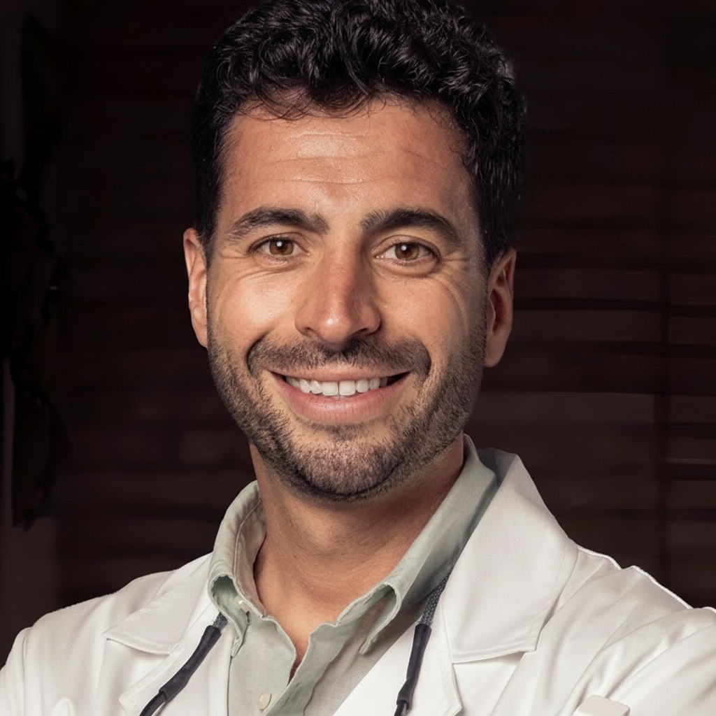
Ignacio Pedrinaci Peñalver centra su práctica en Cirugía Oral e Implantología, con la cirugía guiada como eje de su trabajo. Ejerce en Madrid. Cursó sus estudios de Odontología en Universidad de Granada en 2018.
Con el tiempo, ha sumado formación específica en periodoncia. Su título de posgrado más reciente es el Máster en Periodoncia e Implantología (Universidad Complutense de Madrid, 2020).
Su aportación al colectivo: imparte 2 formaciones — entre ellas «Implantología Digital Contemporánea (3ª edición)» y «Straumann Corporate Forum 2026 · Implantología basada en la evidencia» — en Madrid - España y Riyadh Air Metropolitano, Madrid y firma 35 publicaciones científicas en revistas como Clinical Implant Dentistry and Related Research y The Journal of Prosthetic Dentistry.
Explora su trayectoria
Máster en Periodoncia e Implantología
Máster · Universidad Complutense de Madrid · Madrid, España
Experto en Clínica Periodontal
Experto · Universidad Complutense de Madrid · Madrid, España
Máster Universitario en Ciencias Odontológicas
Máster · Universidad Complutense de Madrid · Madrid, España
Grado / Licenciado en Odontología
Grado · Universidad de Granada · Granada, España
Sin experiencia registrada
Sin actividad docente registrada
Enero a abril de 2027 · Módulos I-IV · Madrid - España · Presencial
Odontología Digital · Cirugía Oral e Implantología
22 de octubre de 2026 · Riyadh Air Metropolitano, Madrid · Presencial
Cirugía y Periodoncia · Implantología
Clinical Implant Dentistry and Related Research
★ Alto impacto · Q1 · IF 3.62Autores · Algirdas Puisys, Samuel Akhondi, Egle Vindasiute‐Narbute, Tadas Zvirblis, German O. Gallucci, Ignacio Pedrinaci
Objectives To evaluate the efficacy of peri‐implant reconstructive therapy using autogenous tuberosity bone grafts, with or without adjunctive electrolytic therapy, as part of surgical treatment for peri‐implantitis. Methods This randomized controlled clinical trial included 31 patients diagnosed with peri‐implantitis. Participants were assigned to either a control group receiving mechanical debridement and bone grafting or a test group undergoing additional electrolytic cleaning (EC). Clinical parameters assessed included probing pocket depth (PPD) at mesial, buccal, distal, and lingual sites, gingival index (GI), and bleeding on probing (BOP), with measurements taken at baseline and at 12 months post‐treatment. Radiographic assessments were conducted at baseline and at the 1‐year follow‐up to evaluate bone defect fill at mesial and distal sites. Intraoperative bone fill was also assessed at mesial, buccal, distal, and lingual sites on the day of surgery and 6 months post‐surgery. Results Both groups exhibited statistically significant PPD reductions at all sites and improvements in GI and BOP compared to baseline. No statistically significant differences between groups were found for these clinical parameters. Radiographically, the test group demonstrated significantly greater mean bone defect fill at the mesial site compared to the control group, with gains of 4.6 mm (±1.10 mm) versus 2.5 mm (±1.77 mm), respectively ( p = 0.002). Intraoperatively, the distal site showed a mean gain of 4.7 mm (±1.41 mm) in the test group and 3.8 mm (±1.72 mm) in the control group after 6 months; however, this difference was not statistically significant ( p = 0.069). Suppuration resolved completely in both groups after 1 year ( p < 0.001), with no significant differences between groups. Conclusions Peri‐implant reconstructive therapy using autogenous bone grafts led to statistically significant clinical and radiographic improvements in both groups. The addition of electrolytic cleaning did not result in significant differences. The absence of histological data, which could not be obtained in this human trial, remains a limitation for confirming re‐osseointegration.
The Journal of Prosthetic Dentistry
★ Alto impacto · Q1 · IF 4.8Autores · Emilio Couso-Queiruga, Ignacio Pedrinaci, Gustavo Avila-Ortiz, Vivianne Chappuis, Eliane Porto Barboza, Rodrigo Lima Petersen, Clemens Raabe, Diogo Moreira Rodrigues
STATEMENT OF PROBLEM: Information regarding the influence of local phenotypical features in the context of immediate implant placement (IIP) in the anterior maxillary region is sparse. PURPOSE: The purpose of this clinical study was to characterize key phenotypical and anatomical characteristics of the anterior maxilla related to the feasibility of virtual IIP. MATERIAL AND METHODS: Cone beam computed tomography (CBCT) scans acquired from adult participants were used for virtual implant placement and to measure periodontal phenotypical dimensions (buccal and palatal gingival (GT) and bone thickness (BT)), the buccal gap (BG) at different apico-coronal levels, and anatomical variables nasopalatine canal and ramifications, nasal cavity, maxillary sinus). The study sample was comprised of 330 maxillary anterior teeth. Two different immediate implant modalities were examined: cingulum emergence plan (CEP) and incisal edge emergence plan (IEP). RESULTS: A total of 660 implants were virtually placed. The mean periodontal phenotypical dimensions showed variability between and within individuals depending on the apico-coronal level, tooth type, and implant placement modality. Immediate implant feasibility was 90.1% and 93.6% for the CEP and IEP groups, respectively, and was influenced by tooth type and anatomical variables. BG distance was generally greater at the coronal aspect and in the CEP. Thick bone and gingiva (≥1 mm) were observed in 15.2% and 89.3% of the sites, respectively. A minimum of 2 mm of apical bone availability to achieve primary stability was observed in 88.8% and 91.2% of the sites in the CEP and IEP groups, respectively. CONCLUSIONS: This study highlights the variability in periodontal phenotypical and local anatomical features at anterior maxillary sites. These observations underscore the importance of recognizing such variations that should be identified and considered during the planning and execution of therapy.
Journal of Esthetic and Restorative Dentistry
★ Alto impacto · Q1 · IF 5Autores · German O. Gallucci, Lorenzo Tavelli, Daniel Buser, Ignacio Pedrinaci, Ronald E. Jung, Kevser Pala
Objective Long‐term stability of peri‐implant hard and soft tissues is a primary goal of implant treatment. Studies have shown that peri‐implant tissue stability is influenced by factors such as the implant characteristics, implant‐abutment interface, abutment type/length, and prosthetic design. To enhance tissue stability while offering clinical versatility, this case report utilizes a dual‐hybrid approach. This combines (1) a hybrid implant surface design, combining a micro‐rough and machined surface, and (2) a hybrid connection where one implant connection allows for two different implant‐abutment interfaces (matching platform/non‐matching with platform switching). Clinical Considerations A patient presented with two posterior teeth deemed “hopeless” due to periodontal and endodontic pathologies. Following extractions, immediate implant placements were performed. Tissue‐level implants were placed subcrestally and restored with non‐matching abutments. The 3‐year follow‐up examination showed stable marginal bone levels maintained at the implant shoulder. Conclusions This dual‐hybrid approach represents an alternative strategy for supporting peri‐implant tissue stability. It underscores the importance of versatile implant‐abutment connections that preserve tissues while allowing future restorative modifications without compromising the implant. This method retains the option to transition to a matching abutment if clinically indicated in the future, for example if any pathologies lead to changes in the peri‐implant tissue levels. Clinical Significance The presented dual‐hybrid approach represents a treatment strategy designed to support peri‐implant tissue stability while maintaining the flexibility to transition to a matching abutment when clinically indicated. Given the prevalence of peri‐implantitis reported in the literature, treatment concepts that not only aim to reduce the risk of disease onset but also facilitate effective reintervention without compromising the implant are of critical importance.
International Journal of Computerized Dentistry
Q3 · IF 1.5Autores · Ignacio Pedrinaci, Amirali Nasseri, Javier Calatrava, Emilio Couso-Queiruga, William V. Giannobile, German O. Gallucci, Mariano Sanz
Coautores Doctor&Cols · Javier Calatrava Serrano de Haro
AIM: The primary aim of the present in vitro study was to compare methods for generating 3D-printed replicas through virtual segmentation, utilizing artificial intelligence (AI) or manual processes, by assessing accuracy in terms of volumetric and linear discrepancies. The secondary aims were the assessment of time efficiency with both segmentation methods, and the effect of post-processing on 3D-printed replicas. MATERIALS AND METHODS: Thirty teeth were scanned through CBCT, capturing the region of interest from human subjects. DICOM files underwent virtual segmentation through both AI and manual methods. Replicas were fabricated with a stereolithography 3D printer. After surface scanning of pre-processed replicas and extracted teeth, STL files were superimposed to compare linear and volumetric differences using the extracted teeth as the reference. Post-processed replicas were scanned to assess the effect of post-processing on linear and volumetric changes. RESULTS: AI-driven segmentation resulted in statistically significant mean linear and volumetric differences of -0.709 mm (SD 0.491, P 0.001) and -4.70%, respectively. Manual segmentation showed no statistically significant differences in mean linear (-0.463 mm, SD 0.335, P 0.001) and volumetric (-1.20%) measures. Comparing manual and AI-driven segmentations, AI-driven segmentation displayed mean linear and volumetric differences of -0.329 mm (SD 0.566, P = 0.003) and -2.23%, respectively. Additionally, AI segmentation reduced the mean time by 21.8 minutes. When comparing post-processed to pre-processed replicas, there was a volumetric reduction of -4.53% and a mean linear difference of -0.151 mm (SD 0.564, P = 0.042). CONCLUSIONS: Both segmentation methods achieved acceptable accuracy, with manual segmentation slightly more accurate but AI-driven segmentation more time-efficient. Continual improvement in AI offers the potential for increased accuracy, efficiency, and broader application in the future.
Clinical Advances in Periodontics
Q3 · IF 1.2Autores · Ignacio Pedrinaci, Javiera Casas, Samuel Akhondi, Javier Calatrava, David Palombo, Alejandro Lanis, German O. Gallucci
Coautores Doctor&Cols · Javier Calatrava Serrano de Haro
BACKGROUND: Peri-implant soft tissues are usually reduced when using a tissue punch in flapless static computer-assisted implant surgery (s-CAIS). The purpose of this article is to introduce a technique combining a minimally invasive guided roll flap (GRF) with s-CAIS to enhance buccal mucosal thickness and maintain peri-implant soft tissues. METHODS: A surgical template is used to define a crest-centered de-epithelialization window and to guide a palatal semilunar full-thickness pedicle that is rolled into a mid-buccal tunnel and stabilized. Osteotomy and implant placement are performed through the same template; the transmucosal profile is supported by an immediate provisional restoration, a sealing socket abutment, or a stock healing abutment. Clinical and volumetric outcomes were assessed over a 12-month follow-up. RESULTS: At 12 months, all implants demonstrated increased buccal mucosal thickness and adequate keratinized mucosa width. Volumetric analysis confirmed soft tissue volume augmentation compared to baseline. No vertical loss of adjacent papillae was observed. Clinical parameters, including PPD and BOP, remained stable, indicating maintained peri-implant health. CONCLUSIONS: This technique demonstrates the feasibility of combining s-CAIS with specific surgical approaches to potentially maintain or slightly improve peri-implant soft tissues. It offers clinicians an alternative method to the soft tissue punch, so the peri-implant soft tissues may be maintained or even augmented in a minimally invasive way.
The International Journal of Prosthodontics
Q3 · IF 1.419Autores · Egle Vindasiutė-Narbute, Algirdas Puisys, Tadas Zvirblis, Kevser Pala, German O. Gallucci, Ignacio Pedrinaci
OBJECTIVES: The choice of retention mechanism in implant-supported restorations (ISRs) strongly influences biological outcomes, with cement-retained ISRs linked to peri-implant complications due to excess cement. Factors such as crown design, margin location, and cement type affect the extent of residual cement and its clinical implications. MATERIAL & METHODS: This prospective, two-arm, non-randomized clinical trial compared cement remnants of self-adhesive resin (RC) and resin-modified glass ionomer (RMGIC) cement in 30 patients, and 60 ISR. Comparisons were performed on identical pairs of ISR tested within the same patients with a 1-week healing interval. The primary outcome variable was the ratio of cement-covered area to total crown area. Areas were analyzed ex-situ by standardized digital image analysis. Statistical analyses included descriptive statistics, Wilcoxon Signed Rank and Kruskal-Wallis tests. RESULTS: All specimens showed remnants of residual cement. In the crown-abutment interface, residual cement was detected on all four crown surfaces and for both types of cement, with total relative cement-covered areas ranging from 0.3% to 11.7% and average values between 1.5% (± 0.7%) and 2.0%. (± 2.4%). RMGIC resulted in higher cement residues than RC in all surfaces except the buccal surface, with differences reaching statistical significance in mesial sites (p=0.007). Other surface comparisons showed no statistical significance. CONCLUSIONS: RMGIC resulted in higher residue areas than RC. Within the limitations of this study, cement remnants appear to be unavoidable through standard clinical procedures, emphasizing the need for case-specific choice of the retention mechanism for ISRs. Screw-retained ISRs present a viable alternative.
Journal of Esthetic and Restorative Dentistry
★ Alto impacto · Q1 · IF 5Autores · Alejandro Lanis, Samuel Akhondi, Ignacio Pedrinaci, Lorenzo Tavelli, Algirdas Puisys
Objective To explore the influence of abutment selection on 3D implant positioning, emphasizing the synergy between surgical and prosthetic considerations for achieving predictable long‐term outcomes in implant‐supported restorations. Main Considerations Implant dentistry has transitioned from a purely surgical approach to a prosthetically driven methodology that prioritizes implant‐supported restoration (ISR). This shift has been bolstered by advancements in digital technologies and abutment designs, which allow for more precise implant positioning and better management of biological, mechanical, and esthetic outcomes. The selection of appropriate abutments plays a pivotal role in optimizing the 3D implant position, influencing peri‐implant tissue stability and the overall success of the restoration. This manuscript explores into the biorestorative concept, highlighting how virtual planning can preemptively assess abutment configurations and their interactions with surrounding tissues, guiding implant placement to achieve desired results. Clinical Significance The integration of digital planning and strategic abutment selection prior to implant placement ensures optimal 3D implant positioning respecting fundamental biological and prosthetic parameters. This approach minimizes complications, improves long‐term tissue stability, and enhances patient outcomes by aligning surgical procedures with the specific prosthetic needs.
Journal of Esthetic and Restorative Dentistry
★ Alto impacto · Q1 · IF 5Autores · Ignacio Pedrinaci, Javier Calatrava, Manuel Toledano‐Osorio, Na Zhao, Alejandro Lanis, Mariano Sanz
Coautores Doctor&Cols · Javier Calatrava Serrano de Haro
Objectives To demonstrate how contemporary digitally driven workflow can enhance outcomes for complex esthetic dental cases, focusing on three distinct clinical scenarios involving implant placement, esthetic crown lengthening, and tooth autotransplantation (ATT). Overview Three multidisciplinary clinical cases demonstrate our contemporary digital workflows, integrating diagnosis, treatment planning, patient communication, and guided execution. The first case involves replacing two anterior central incisors using digital planning, guided surgery, prefabricated customized healing abutments, and a digitally driven restorative process. The second case showcases an esthetic crown‐lengthening procedure, where Multifunctional Anatomical Prototypes (MAPs) serve as both mock‐up and surgical guides, enhancing patient communication on expected outcomes and ensuring precise tissue management to prevent soft tissue rebound. The final case features a tooth ATT, where virtual surgical planning and 3D‐printed tooth replica and guides ensure predictability in the therapeutic outcome. These digitally enabled strategies underscore the predictability and customization achievable with contemporary dental technology. Conclusions Dental treatments in the esthetic zone require meticulous planning and precise execution to achieve controlled results that ensure patient satisfaction and long‐term stability. Contemporary Digital Dentistry enhances predictability, creating a pathway that leads to success.
Medicina Oral Patología Oral y Cirugia Bucal
Q2 · IF 2.2Autores · JF. Peña-Cardelles, J. Markovic, S. Akhondi, I. Pedrinaci, A. Lanis, GO. Gallucci
BACKGROUND: A concern associated with implant placement is the potential occurrence of neurovascular lesions and subsequent development of sensory alterations in patients undergoing implant placements. The objective of this review is to evaluate the incidence of neurosensory alterations based on the proximity between the implant and the mandibular canal Material and Methods: A systematic review was conducted in MEDLINE, Web of Science, and Scopus. Studies with common variables were selected to conduct a meta-analysis. The patient classification was based on the mandibular canal-implant distance. Neurosensory alteration percentages were calculated for each study and group. RESULTS: The findings indicate significant correlations between implant placement proximity and neurosensory risks. The incidence of neurosensory alterations in patients with implants placed at a distance ≥ 2 mm from the mandibular canal was 0%. Similarly, for distances between 1-2 mm from the mandibular canal, the incidence remained at 0%. However, for implants placed at a distance of 0-1 mm from the mandibular canal, the incidence of neurosensory alterations was 68%. Additionally, patients with implants that intruded into the canal had an incidence of 53% in the development of neurosensorial alterations. CONCLUSIONS: A distance of 1 mm from the mandibular canal might be safe. Implants placed at a distance less than 1 mm from the mandibular canal exhibit neurosensory alterations. Clinicians should be aware of the potential risk of nerve injury and adopt appropriate precautions, including meticulous preoperative planning and three-dimensional radiographic images.
Clinical Implant Dentistry and Related Research
★ Alto impacto · Q1 · IF 3.62Autores · Ignacio Pedrinaci, Betty Ben Dor, Dominique Rousson, Alejandro Lanis, Javier Sanz‐Esporrin, Kevser Pala, German O. Gallucci, Adam Hamilton
Introduction Using mini implants as transitional implants (TIs) for complete arch implant‐supported rehabilitations may overcome limitations associated with mucosa‐supported surgical guides and facilitate immediate fixed provisionalization. This study aimed to assess the success of TIs in supporting surgical guides for implant placement and fixed provisional prostheses. Methods Patients who received TIs between 2012 and 2023 for a complete arch implant‐supported prosthesis were evaluated retrospectively. Patient demographic data, TI functionality in supporting a surgical guide and supporting a complete arch provisional prosthesis, and dates of TI placement and regular implant placement were collected. Descriptive statistics were used to determine the survival rate and success rate for TIs. Results Twenty‐six patients, 35 jaws, 136 TIs, and 216 regular implants were included. The survival rate of TIs was 74.26%; however, the use of TIs yielded success in 97% of jaws for supporting a surgical guide and a fixed complete‐arch provisional prosthesis throughout the complete provisional phase. An average of 4 TIs per maxilla and 3 TIs per mandible supported surgical guides. Thirty‐five provisional prostheses were placed on an average of 4 TIs in the maxilla and 3 TIs in the mandible. Thirty‐four provisional prostheses were successfully supported by TIs and regular implants until final restoration delivery. The survival of regular implants placed in conjunction with the use of TIs was 98%. Conclusions Using TIs to support a surgical guide and provisional prosthesis may be a predictable approach with a high success rate. All surgical guides planned to be supported on TIs were successful. Despite premature loss or replacement of TIs, this approach was able to support most provisional prostheses until the regular implants could be loaded.
Periodoncia Clínica (SEPA) nº33 · Monográfico 'Impresión 3D en la consulta dental'
Autores · Pedrinaci I, Finelle G, Gallucci GO
La planificación digital y la impresión 3D han transformado la implantología contemporánea al permitir una integración precisa entre el enfoque quirúrgico y protético bajo el concepto biorestaurador (BR). Se describe el tratamiento de una paciente de 43 años con fractura horizontal del 25: extracción atraumática, colocación inmediata de implante mediante cirugía guiada (sCAIS), preservación alveolar con xenoinjerto y sellado con un pilar de cicatrización personalizado (Sealing Socket Abutment) diseñado a partir de una réplica 3D del resto radicular. El flujo incluyó CBCT e IOS, segmentación con inteligencia artificial, planificación protética 3D y fabricación interna de la férula quirúrgica y del SSA.
Clinical Oral Implants Research
★ Alto impacto · Q1 · IF 4.3Autores · Krisztina Mikulás, Xinyi Qian, Péter Tajti, Gergely Agócs, German O. Gallucci, Ignacio Pedrinaci, Péter Hermann
Background Accurately replicating the emergence profile (EP) of conditioned soft tissue is pivotal for the success of implant‐supported restorations. In the field of digital technology, various methods have emerged to capture EP. This review aims to critically assess current digital methodologies for capturing peri‐implant EP. Material and Methods Prospective interventional or observational clinical studies focusing on digitally mapping the emergence profile (EP) around single implant‐supported restorations were included. Systematic reviews, in vitro and animal studies, and those not emphasizing EP capture were excluded. A systematic search across four databases (MEDLINE, CENTRAL, Embase, Web of Science) was conducted on 7th August 2024 based on a previously registered protocol (PROSPERO registration number: CRD42023459484). Risk of bias was assessed with RoB 2, ROBINS‐I, and JBI critical appraisal tools. Qualitative and quantitative analyses were carried out. Results Twenty‐four eligible studies were identified, comprising 5 dental techniques, 12 case reports, 1 randomized controlled study, 2 cross‐sectional studies, and 4 cross‐over studies. The studies reported semi‐digital pathways, direct scanning, indirect scanning, coded‐healing abutments, and individualized use of scan bodies. Notably, the direct scanning technique showed considerable soft tissue collapse. Similar results can be achieved with indirect scanning and the conventional method. Conclusions Indirect EP scanning appears as the most promising method for capturing peri‐implant EP. However, a confirmation of this finding requires a quantitative analysis through randomized clinical trials.
Journal of Esthetic and Restorative Dentistry
★ Alto impacto · Q1 · IF 5Autores · Fernando Cebrián, Francisco Carroquino, Ignacio Pedrinaci
Coautores Doctor&Cols · Fernando Cebrián Vicente, Francisco Carroquino Cuevas
Peri-implant soft tissue is considered critical for maintaining peri-implant health, minimizing esthetic and biological complications, and promoting the long-term stability of implant-supported restorations. This article describes a straightforward and minimally invasive technique for augmenting the peri-implant mucosa thickness (MT) in the mandibular posterior area using a rotated pedicled flap (MRPF). The surgical procedure was conducted on sixteen systemically healthy adults (8 males, 8 females) with posterior mandibular partial edentulism and a thin tissue phenotype. Clinical and digital analysis variables demonstrated a peri-implant mucosal thickness increase (2.72 ± 0.69 mm) at 12 months follow-up after the definitive implant-supported restoration delivery. The proposed surgical technique successfully increased the buccal peri-implant MT.
The International Journal of Oral & Maxillofacial Implants
Autores · Samuel Akhondi, Kevser Pala, Ignacio Pedrinaci, German O. Gallucci
This technical note serves as a practical guide for clinicians aiming to provide solutions to optimize the precision of static computer-assisted implant surgery (sCAIS) and the success of implant placement in free-end situations. Five methods are introduced for enhancing surgical guide stabilization and improving the accuracy of implant placement in situations lacking stabilizing adjacent teeth. These techniques include the use of keratinized soft tissue, implant-borne stabilizers, lateral fixation pins, transitional implants, and digital bone segmentation for guide support. Each method is evaluated for its potential to address the specific challenges faced in sCAIS, aiming to contribute to the field through practical solutions for clinicians.
Journal of Esthetic and Restorative Dentistry
★ Alto impacto · Q1 · IF 5Autores · Ignacio Pedrinaci, Adam Hamilton, Alejandro Lanis, Mariano Sanz, German O. Gallucci
Objective This study aims to present the bio‐restorative approach in implant dentistry, which combines biological and restorative concepts through digital planning. This concept combines periodontal, surgical, and prosthetic variables, aiming to reduce patient morbidity while achieving satisfactory esthetic and functional outcomes in implant‐supported restorations in the long term. Overview Implant dentistry evolved from a primarily surgical to a recent prosthetically driven approach. This evolution was partly due to advancements in bone reconstructive techniques and an increased demand for esthetic outcomes. Recently, digital planning has introduced a new paradigm that allows for the full integration of both approaches. The bio‐restorative concept considers functional, esthetic, and biological variables in a virtual planning environment. This is achieved through the simultaneous digital assessment of (A) anatomical site characteristics and (B) implant restorative variables. These variables include digital tooth arrangement, soft–hard tissue conditions, implant variables, supra‐platform components, and a surgical plan that respects or modifies peri‐implant phenotype. Conclusions The bio‐restorative concept is intended to improve contemporary implant dentistry by integrating updated biological and prosthetic notions through digital planning. Adopting this paradigm has the potential to redefine the standards in implant dentistry, fostering a holistic and patient‐centered approach. Clinical Considerations It enhances patient and clinician satisfaction through more efficient and less invasive procedures. Significantly, it improves predictability, leading to successful implant‐supported restorations in the long term.
International Journal of Periodontics & Restorative Dentistry
Autores · Ignacio Pedrinaci, German O. Gallucci, Alejandro Lanis, Bernard Friedland, Kevser Pala, Adam Hamilton
Computer-assisted implant planning allows for a comprehensive treatment plan by combining radiographic data provided by CBCT with surface optical scan data that includes the patient's intraoral situation and the intended restorative planning. Integrating a tailored restorative de-sign with the patient's anatomical conditions through virtual implant planning allows for an ideal biorestorative treatment planning that maximizes biologic, functional, and esthetic outcomes. This article discusses dataset registration techniques that combine radiographic CBCT data with re-storative information as the main path to create a virtual patient. The described techniques include the use of removable radiographic templates with radiopaque markers, the dual scan technique, and direct digital file registration of intraoral scans using anatomical references. Depending on the individual clinical situation, different factors must be considered to appropriately select methods that achieve an optimal registration of diverse datasets. An inherent challenge lies in the presence of scattering artifacts in CBCT scans. Two approaches are proposed for these situations: the use of chairside-fabricated composite resin markers or adhesive spot-markers fabricated for use with computed tomography scans. Both techniques exhibit limitations that must be considered. Further approaches should be developed for situations involving scattering in CBCT scans.
Journal of Esthetic and Restorative Dentistry
★ Alto impacto · Q1 · IF 5Autores · Jovana Markovic, Juan Francisco Peña‐Cardelles, Ignacio Pedrinaci, Adam Hamilton, German O. Gallucci, Alejandro Lanis
Objective To provide technical and clinical recommendations for implementing a digital workflow in Static Computer‐Aided Implant Surgery in the anterior maxilla. Clinical Considerations An optimal 3D implant position is crucial for achieving satisfying results in implant rehabilitation in the esthetic area. Due to its complexity, implant placement in the esthetic zone should be executed with precision and predictability. Static Computer‐Aided Implant Surgery requires thorough planning and detailed attention to every step of the digital workflow protocol. Conclusions Implant positioning in the esthetic zone using Static Computer‐Aided Implant Surgery is a technique‐sensitive procedure that requires precise execution of each step. This approach ensures accurate prosthetically driven 3D implant placement and prevents potential errors that could lead to inaccurate positioning. Clinical Significance The proper implementation of Static Computer‐Aided Implant Surgery may increase the level of agreement between the planned and definitive implant 3D positions in the esthetic zone, thus enhancing the esthetic outcomes of implant rehabilitation.
AJO-DO Clinical Companion
Autores · Christopher Burns, Elli Anna Kotsailidi, Fares Alshuraim, Shaahin Dadjoo, Konstantina Tzouma, Ignacio Pedrinaci, Paul Emile Rossouw, Dimitrios Michelogiannakis
BACKGROUND: Measuring the ability of SARS-CoV-2 antibodies to neutralize live viruses remains an effective approach to quantify the level of protection of individuals. We assessed the neutralization activity against the ancestral SARS-CoV-2, Delta, Omicron BA.1, BA.2, BA.2.12.1, BA.4 and BA.5 strains, in 280 vaccinated restaurant/bar, grocery and hardware store workers in Québec, Canada. METHODS: Participants were recruited during the emergence of Omicron BA.1 variant. The neutralizing activity of participant sera was assessed by microneutralization assay. RESULTS: Serum neutralizing antibody (NtAb) titers of all participants against the ancestral SARS-CoV-2 strain were comparable with those against Delta variant (ranges of titers 10-2032 and 10-2560, respectively), however, their response was significantly reduced against Omicron BA.1, BA2, BA.2.12.1, BA.4 and BA.5 (10-1016, 10-1016, 10-320, 10-80 and 10-254, respectively). Individuals who received 2 doses of vaccine had significantly reduced NtAb titers against all SARS-CoV-2 strains compared to those infected and then vaccinated (≥1 dose), vaccinated (≥2 doses) and then infected, or those who received 3 doses of vaccine. Participants vaccinated with 2 or 3 doses of vaccine and then infected had the highest NtAb titers against all SARS-CoV-2 strains tested. CONCLUSION: We assessed for the first time the NtAb response in food and retail workers. We found that vaccination prior to the emergence of Omicron BA.1 was associated with higher neutralizing activity against pre-Omicron variants, suggesting the importance of updating vaccines to increase antibody response against new SARS-CoV-2 variants. Vaccination followed by infection was associated with higher neutralizing activity against all SARS-CoV-2 strains tested.
Journal of Dentistry
★ Alto impacto · Q1 · IF 4.94Autores · Ignacio Pedrinaci, Javier Calatrava, Emilio Couso-Queiruga, Juan del Rosal Bethencourt, Ignacio Sanz-Sanchez, German O. Gallucci, Mariano Sanz
Coautores Doctor&Cols · Javier Calatrava Serrano de Haro
OBJECTIVES: Digital protocols and bioactive materials may reduce complications and improve tooth autotransplantation (ATT) success and survival rates. This prospective study assesses the performance of a fully digital autotransplantation protocol of close-apex molars with the adjunctive application of Enamel Matrix Derivatives (EMD). METHODS: Twelve adult patients with 13 hopeless molar teeth were replaced with autotransplantation of closed apex third molars. Outcomes, including success and survival rates, clinical, endodontic, radiographic, patient-reported outcome measures (PROMs), and digital image assessments, were conducted over a two-year follow-up period. RESULTS: Survival and success rates were 100% and 91.2%, respectively, with no progressive inflammatory or replacement root resorption (ankylosis) except for one tooth presenting radiographic furcation involvement. A significant probing depth reduction of 2.4 ± 2.58 mm and CAL gains of 2.8 ± 3.03 mm were observed in transplanted teeth compared to the hopeless receptor teeth. Radiographic bone levels remained stable throughout the study period (-0.37 ± 0.66 mm), and digital image assessments showed minimal alveolar ridge width changes (-0.32 to -0.7 mm) and gingival margin changes (-0.95 to -1.27 mm) from baseline to last visit. PROMs indicated very high patient satisfaction. CONCLUSION: The use of a digital ATT protocol with adjunctive use of EMD in closed-apex third molars demonstrated promising short-term high success and survival rates. Additionally, this type of therapy adequately preserves the dimensions of the alveolar ridge in the receptor site. CLINICAL SIGNIFICANCE: This is the first prospective clinical study examining the effect of a digital tooth autotransplantation protocol combined with the application of EMD. It demonstrates that this approach is an effective treatment for replacing hopeless teeth and also validates the digital assessment of ATT alveolar ridge preservation at the recipient site.
Clinical Implant Dentistry and Related Research
★ Alto impacto · Q1 · IF 3.62Autores · Algirdas Puisys, Egle Vindasiute‐Narbute, Dainius Razukevicius, Samuel Akhondi, German O. Gallucci, Ignacio Pedrinaci
Objectives This study aimed to compare the efficacy of two techniques—acellular dermal matrix (ADM) grafting and tenting technique (TT)—for soft tissue height (STH) augmentation simultaneous to implant placement to minimize peri‐implant crestal bone level (CBL) changes. Methods Forty patients with a healed single mandibular posterior edentulous site with a thin soft tissue phenotype were enrolled. Twenty patients received simultaneously to implant placement ADM grafting, while the others received submerged healing abutment (TT). Clinical peri‐implant soft tissue height and radiographic CBL changes were measured at restoration delivery and 1‐year follow‐up. Results Both techniques effectively increased soft tissue thickness, resulting in a final average STH of 3.4 ± 0.5 mm after augmentation. On average, soft tissue increased by 1.6 ± 0.5 mm in group ADM and by 1.8 ± 0.4 mm in group TT after augmentation. In Group ADM, mesial CBL decreased from 0.4 ± 0.3 mm to 0.1 ± 0.2 mm, and distal CBL decreased from 0.5 ± 0.3 mm to 0.2 ± 0.3 mm over 1 year. In Group TT, mesial CBL remained stable at 0.3 ± 0.2 mm, while distal CBL reduced slightly from 0.5 ± 0.5 mm to 0.3 ± 0.2 mm. Both groups showed minimal changes in CBL, indicating great stability ( p mesial = 0.003, p distal = 0.004). TT was particularly effective in preventing mesial bone loss ( p mesial = 0.019). The mesial CBL changes significantly differed between groups ( p = 0.019), and not significantly at distal sites ( p = 0.944). Neither treatment exhibited significant bone remodeling below the implant shoulder. Conclusion This study suggests that both techniques were successful in STH augmentation, and they may effectively reduce peri‐implant crestal bone level changes, with TT being slightly superior. TT was more prone to post‐surgical complications. This RCT was not registered before participant recruitment and randomization.
The International Journal of Periodontics & Restorative Dentistry
Autores · Juan Francisco Peña-Cardelles, Fernando Núñez Díaz, Elli Kotina, Ignacio Pedrinaci, Alejandro Lanis, German O. Gallucci
Introduction: Maxillary sinus floor augmentation is a procedure known for its long-term success and predictable outcomes. However, the perforation of the Schneiderian membrane remains the most common complication associated with this procedure. Objective: This systematic review aims to determine the presence of complications during maxillary sinus floor augmentation procedures using CAD-CAM surgical templates. Material and methods: An electronic search was carried out in MEDLINE (via PubMed), Web of Science, and Scopus. A descriptive analysis of the data was performed. Studies that have performed lateral sinus floor augmentation were included in the inclusion criteria. The CAD-CAM surgical template design and the intraoperative complications were registered. Results: A total of 13 studies were included. Seven were case reports, four were case series, and two were randomized clinical trials. A total of 94 lateral SFA procedures were included (84 using CAD-CAM templates and 10 without using templates). Three of the 84 maxillary sinus floor augmentation procedures using a CAD-CAM template presented intraoperative complications. Conclusions: Maxillary sinus floor augmentation performed by using CAD-CAM surgical templates could be related to low rates of complications, however, due to the heterogeneity of the articles included, more standardized studies are needed to confirm these outcomes.
The International Journal of Prosthodontics
Q3 · IF 1.419Autores · Elli Kotina, Adam Hamilton, Jason D Lee, Sang J Lee, Peter C Grieco, Ignacio Pedrinaci, Neil T Griseto, German O Gallucci
Traditionally, metal-ceramics, metal-reinforced acrylics, and-more recently-full-contour or layered zirconia have been the materials of choice for definitive fixed implant-supported rehabilitations. Polymethyl methacrylate (PMMA) is commonly used in implant dentistry for the fabrication of implant-supported interim prostheses and as milled or 3D-printed prototypes. This article describes a novel protocol to prosthetically restore a completely edentulous patient following a digital workflow, with fixed, screw-retained, implant-supported prostheses fabricated from CAD/CAM milled PMMA, with no metal substructure. After a 2-year follow-up in terms of esthetics, phonetics, function, and biologic tissue response, the outcome remains functional and free of mechanical, biomechanical, or biologic complications. The aim of this article is to illustrate the feasibility of using milled PMMA as a viable definitive prosthetic material for the fixed implant rehabilitation of edentulous patients.
Journal of Dentistry
★ Alto impacto · Q1 · IF 4.94Autores · Algirdas Puisys, Egle Vindasiute-Narbute, Danius Razukevicius, Samuel Akhondi, German O. Gallucci, Ignacio Pedrinaci
OBJECTIVE: To evaluate the 10-year influence of soft tissue height (STH) on crestal bone level changes (CBC) in bone-level implants with non-matching internal conical connections. MATERIAL & METHODS: From the initial 97 patients, 59 (19 men, 40 women, age 55.86 ± 9.5 years) returned for the recall visit. Based on baseline STH, they were categorized into T1 (thin STH ≤2 mm, n = 33), T2 (thin STH augmented with allogenic tissue matrix (ATM), n = 32), and C (thick STH >2 mm, n = 32). Implants were placed in the posterior mandible using a one-stage approach and received single screw-retained restorations. Clinical (PPD, BOP, PI) and radiographic examinations were conducted after 10 years, with CBC calculated mesial and distal to each implant. RESULTS: After 10 years, implants in surgically thickened (T2) or naturally thick STH (C) showed bone gains of 0.57 ± 0.55 mm and 0.56 ± 0.40 mm, respectively (p < 0.0001) shifting from an initial CBC of -0.21 ± 0.33 mm to 0.36 ± 0.29 mm in the thick STH group and -0.2 ± 0.35 mm to 0.37 ± 0.29 mm in the surgically thickened STH group. Implants in naturally thin STH yielded a non-significant trend of bone loss (-0.12 ± 0.41 mm; p > 0.05). CONCLUSIONS: Implants in thin STH (≤2 mm) exhibited greater CBC over the study period. Significant bone gains were observed in thick STH cases, indicating that naturally thick STH or STH augmentation with ATM may contribute to maintain CBC in long-term around implants. CLINICAL SIGNIFICANCE: This is the first long-term follow-up study suggesting that adequate soft tissue height around implants helps maintain stable peri‑implant bone levels. While tissue thickness plays a key role, other factors also interact with peri‑implant tissue height to sustain crestal bone stability over time.
Journal of Esthetic and Restorative Dentistry
★ Alto impacto · Q1 · IF 5Autores · Ignacio Pedrinaci, Javier Calatrava, Juan Flores, Adam Hamilton, German O. Gallucci, Mariano Sanz
Coautores Doctor&Cols · Javier Calatrava Serrano de Haro
Objective To describe a strategy using digital technologies for improving the diagnosis, treatment planning, and surgical execution of patients with excessive gingival display (EGD) due to altered passive eruption (APE). Clinical Considerations An important component for successful patient's management is to fulfill their esthetic expectations whilst delivering predictable and long‐term therapeutic outcomes. To achieve this goal in patients with excessive gingival display due to altered passive eruption, it is essential to perform an accurate diagnosis and to communicate to the patient the expected customized results using digital technologies. Computer‐aided designed and manufactured multifunctional anatomical prototypes (MAPs) may contribute to these purposes. Additionally, they can guide the surgical crown lengthening procedure or serve as a reference during the surgical guide fabrication providing information of the required anatomical landmarks. Conclusions This novel strategy protocol for diagnosis, communication, and treatment management of patients with excessive gingival display follows functional and biological principles within the frame of a digital workflow, which improves the diagnostic capabilities, enhances communication, and guides the surgical treatment as shown in the 12 months follow‐up of the reported case. Clinical Significance Developing a virtual patient by combining multiple digital data sets including cone‐beam computed tomography (CBCT), intra‐oral scans and digital photography, supports the clinician and the patient to achieve a comprehensive diagnosis and to better communicate the expected results to the patient. Furthermore, this digital treatment exercise based on anatomical and biological principles will facilitate the surgical precision and the achievement of successful outcomes, thus fulfilling the patient needs and expectations.
International Journal of Environmental Research and Public Health
★ Alto impacto · Q1 · IF 3.251Autores · Dániel Végh, Bulcsú Bencze, Dorottya Banyai, Adam Vegh, Noémi Rózsa, Csaba Nagy Dobó, Zita Biczo, Gabor Kammerhofer, Marta Ujpal, Leonardo Díaz Agurto, Ignacio Pedrinaci, Juan Francisco Peña Cardelles, Gabriel Leonardo Magrin, Ninad Milind Padhye, Laura Mente, Michael Payer, Peter Hermann
Diabetes mellitus has become a worldwide epidemic and is frequently accompanied by a number of complications proportional to the duration of hyperglycemia. The aim of this narrative review is to assess the most up-to-date guidelines on DM provided by both diabetes and dental associations. Furthermore, to gather evidence on the uni/bidirectional relationships of elevated HbA1c levels on dental surgery, implantology, bone augmentation, and periodontology and to demonstrate the importance of measuring HbA1c levels before invasive dental treatments. HbA1c and blood glucose measurements are a minimally invasive method for preventing complications in diabetes mellitus. The authors conducted a literature review to determine which oral conditions are affected by diabetes mellitus. MEDLINE served as a source with the use of a specific search key. Regarding oral complications of diabetes, prevention is the most vital factor. With this publication, we hope to assist physicians and dentists to make prompt diagnoses and to help in recognizing various oral manifestations of diabetes and follow the existing guidelines.
International Dental Journal
★ Alto impacto · Q1 · IF 3.61Autores · Peña-Cardelles JF, Pedrinaci I, Kotina E, Lanis A, Salgado-Peralvo AO
Coautores Doctor&Cols · Orion Salgado
Este artículo analiza el potencial de la cirugía implantológica asistida por ordenador estática como herramienta para optimizar la planificación y ejecución de los tratamientos implantarios. A partir de un enfoque clínico y tecnológico, explora cómo la mayor precisión quirúrgica, la reducción del trauma tisular y la mejora de la previsibilidad podrían contribuir a disminuir complicaciones infecciosas y, potencialmente, la necesidad de antimicrobianos. Su interés clínico radica en situar la odontología digital dentro de las estrategias de uso prudente de antibióticos y de lucha frente a la resistencia antimicrobiana.
Journal of Esthetic and Restorative Dentistry
★ Alto impacto · Q1 · IF 5Autores · Alejandro Lanis, German Gallucci, Ignacio Pedrinaci
Objective To demonstrate the use of a complete digital workflow for a full mouth rehabilitation in a severely worn dentition. Clinical Considerations The present case report successfully rehabilitated a full‐mouth case of a severely worn dentition based on the use of digital technologies, making the diagnosis and treatment process faster, accurate and less expensive. A long‐lasting esthetic and functional result are showed after 30‐months follow up. Conclusions An appropriate knowledge on dental erosion and oral rehabilitation, combined with a digital dentistry approach could lead the clinician to deliver a fast, accurate and predictable noninvasive restorative treatment in cases like the one described. Clinical Significance Bruxism‐based severely worn dentition is being found more often in population. In this situation, a detailed diagnosis and tailored treatment are mandatory to obtain a predictable treatment outcome. In this sense, the development of adhesive dentistry, new restorative materials and the incorporation of digital technologies can create a predictable synergy to rehabilitate these types of patients with a modern and less invasive approach.
Clinical Oral Implants Research
★ Alto impacto · Q1 · IF 4.3Autores · Ignacio Pedrinaci, Teresa Chanting Sun, Mariano Sanz‐Alonso, Javier Sanz‐Esporrin, Adam Hamilton, German O. Gallucci
Objective This study aims to report the implant survival rate of dental implants of partially dentate patients in the anterior mandible and the potential risk indicators for implant failure. Materials and Methods Patients with implant‐supported restorations of single or multiple teeth in the anterior mandible restored with fixed partial implant‐supported restorations were evaluated. Patient demographic data, implant placement timing, and loading protocol, biological and/or technical complications at the time of the last clinical and radiographic follow‐up visit were registered. Survival rate, success rate, and potential risk indicators for implant failure were calculated. Results A total of 108 patients and 186 implants with a mean follow‐up period of 5.48 years (0.1–11.34 years) were included. The 11.3‐year cumulative survival rate was 90.9%. Immediate implant placement (OR = 2.75) ( p = .08) and immediate implant loading (OR = 8.8) ( p = .02*) indicated a higher risk of failure than late implant placement or loading. When combining both categories (type 1A), an OR = 10.59 ( p = .04*) for implant failure was found compared to category 4C. Implants placed following static—computer‐assisted implant surgery (S‐CAIS) showed less risk of failure compared to freehand implant placement (OR = 0.18; 95% CI: 0.02–1.37) ( p = .09). Conclusions The survival rate of implants placed in the anterior mandible was considerably low (90.9%). S‐CAIS, late placement, and conventional loading are protective factor against implant failure in the anterior mandible.
Journal of Clinical Periodontology
★ Alto impacto · Q1 · IF 5.8Autores · Mario Romandini, Andreina Laforí, Ignacio Pedrinaci, Giacomo Baima, Francesco Ferrarotti, Cristina Lima, Lucrezia Paternó Holtzman, Mario Aimetti, Luca Cordaro, Mariano Sanz
Aim The present multi‐centre randomized clinical trial with 12 months of follow‐up aimed at studying the added effect of sub‐marginal instrumentation before surgical treatment of peri‐implantitis. Materials and Methods Forty‐two patients diagnosed with peri‐implantitis were recruited. After a behavioural intervention phase including oral hygiene instructions, patients were randomized to either receiving supra‐ and sub‐marginal instrumentation on their affected implants (control group: 21 patients and 29 implants) or only supra‐marginal instrumentation (test group: 21 patients and 24 implants), before undergoing surgery. Changes in the deepest probing pocket depth (PPD) with respect to baseline and a composite outcome of treatment success (no implant loss, no bone loss > 0.5 mm, no bleeding or suppuration on probing [BoP/SoP], and PPD ≤ 5 mm) at the 12‐month examination were regarded as the primary outcomes of the trial. Results At the 12‐month examination, changes in the deepest PPD with respect to baseline amounted to −2.96 mm in the control group and to −3.11 mm in the test one (MD = −0.16; SE = 0.56; p = .769), while 21.4% of the implants in the control group and 33.3% in the test group presented treatment success (OR = 1.83; SE = 1.15; p = .338). With the exception of a longer non‐surgical treatment duration in the control group (differences in = −14.29 min; SE = 2.91; p < .001), no other secondary (e.g., soft‐tissue recession, keratinized mucosa height, and bone level changes, as well as BoP, SoP, profuse bleeding and implant loss rates) or exploratory (i.e., early wound healing, aesthetics, surgical and total treatment duration, surgery difficulty, intra‐operative bleeding, and adverse events) outcome demonstrated statistically significant differences between groups. Conclusions The present multi‐centre randomized clinical trial did not demonstrate an added effect of performing sub‐marginal instrumentation 6 weeks before the surgical treatment of peri‐implantitis. Larger clinical trials are however needed to confirm the present findings (Clinicaltrials.gov: NCT03620331).
Periodoncia Clínica (SEPA) nº23 · Monográfico 'Science and practice of tooth autotransplantation'
Autores · Pedrinaci I, Calatrava J, Sanz-Sánchez I, Sanz M
Coautores Doctor&Cols · Javier Calatrava Serrano de Haro
Los autotrasplantes dentales presentan tasas de supervivencia superiores al 90% con seguimientos de más de 20 años, y su éxito depende directamente de la vitalidad del ligamento periodontal del diente donante. Se presenta el caso de un varón de 17 años con un primer molar inferior izquierdo no restaurable, sustituido por el tercer molar del mismo cuadrante mediante un flujo de trabajo completamente digital: CBCT convertido a STL, modelo estereolitográfico mandibular impreso en 3D, réplica del diente donante y férula de cirugía guiada para la osteotomía del alvéolo receptor. Antes de reposicionar el diente se aplicaron proteínas derivadas de la matriz del esmalte (EMD) sobre la superficie radicular para aumentar el potencial regenerativo y reducir las complicaciones del procedimiento.
Forum Implantologicum
Autores · Ignacio Pedrinaci, Neil Griseto, Adam Hamilton, German O. Gallucci
Achieving an ideal esthetic outcome with implant-supported rehabilitation of missing teeth is a challenging procedure, especially in the anterior zone. Numerous clinical variables have been reported to impact the ultimate treatment outcome. Thus, a comprehensive understanding of the anatomic, biologic, surgical, prosthetic and patient-centered factors must be pursued to obtain an optimal and predictable result. This article summarizes most of the variables contributing to esthetic and biological success in implant prosthodontics of the anterior zone and to provide key treatment considerations on the decision-making process of replacing missing anterior teeth with dental implants.
Antibiotics
★ Alto impacto · Q1 · IF 5.77Autores · Ángel-Orión Salgado-Peralvo, Juan‐Francisco Peña‐Cardelles, Naresh Kewalramani, Alvaro Garcia-Sanchez, María‐Victoria Mateos‐Moreno, Eugenio Velasco‐Ortega, Iván Ortiz-García, Álvaro Jiménez-Guerra, Dániel Végh, Ignacio Pedrinaci, Loreto Monsalve-Guil
Coautores Doctor&Cols · Naresh Kewalramani, Orion Salgado
As the population ages, more and more patients with orthopaedic prostheses (OPs) require dental implant treatment. Surveys of dentists and orthopaedic surgeons show that prophylactic antibiotics (PAs) are routinely prescribed with a very high frequency in patients with OPs who are about to undergo dental procedures. The present study aims to determine the need to prescribe prophylactic antibiotic therapy in patients with OPs treated with dental implants to promote their responsible use and reduce the risk of antimicrobial resistance. An electronic search of the MEDLINE database (via PubMed), Web of Science, LILACS, Google Scholar, and OpenGrey was carried out. The criteria used were those described by the PRISMA® Statement. No study investigated the need to prescribe PAs in patients with OPs, so four studies were included on the risk of infections of OPs after dental treatments with varying degrees of invasiveness. There is no evidence to suggest a relationship between dental implant surgeries and an increased risk of OP infection; therefore, PAs in these patients are not justified. However, the recommended doses of PAs in dental implant procedures in healthy patients are the same as those recommended to avoid infections of OPs.
Clinical Oral Implants Research
★ Alto impacto · Q1 · IF 4.3Autores · Mario Romandini, Cristina Lima, Ignacio Pedrinaci, Ana Araoz, Maria Costanza Soldini, Mariano Sanz
Aim To evaluate the prevalence of peri‐implant diseases and to identify risk/protective indicators of peri‐implantitis. Materials and Methods Two hundred and forty randomly selected patients from a university clinic database were invited to participate. Those who accepted, once data from their medical and dental history were collected, were examined clinically and radiographically to assess the prevalence of peri‐implant health and diseases. Peri‐implantitis was defined as the presence of BoP/SoP together with radiographic bone levels (BL) ≧2 mm. An intermediate peri‐implant health category between peri‐implant mucositis and peri‐implantitis was also identified, defined by the presence of BoP/SoP together with 1 mm ≦BL < 2 mm. A multilevel multivariate logistic regression analysis was carried out to identify those factors associated either positively (risk) or negatively (protective) with peri‐implantitis. Results Ninety‐nine patients with a total of 458 dental implants were analyzed. The prevalences of pre‐periimplantitis and of peri‐implantitis were, respectively, 31.3% and 56.6% at patient‐level, while 31.7% and 27.9% at implant level. The following factors were identified as risk indicators for peri‐implantitis: smoking (OR = 3.59; 95% CI: 1.52–8.45), moderate/severe periodontitis (OR = 2.77; 95% CI: 1.20–6.36), <16 remaining teeth (OR = 2.23; 95% CI: 1.05–4.73), plaque (OR = 3.49; 95% CI: 1.13–10.75), implant malposition (too vestibular: OR = 2.85; 95% CI: 1.17–6.93), implant brand (Nobel vs. Straumann: OR = 4.41;95% CI: 1.76–11.09), restoration type (bridge vs. single crown: OR = 2.47; 95% CI: 1.19–5.12), and trauma as reason of tooth loss (vs. caries: OR = 6.51; 95% CI: 1.45–29.26). Conversely, the following factors were identified as protective indicators: interproximal flossing/brushing (OR = 0.27; 95% CI: 0.11–0.68), proton pump inhibitors (OR = 0.08; 95% CI: 0.01–0.90), and anticoagulants (OR = 0.08; 95% CI: 0.01–0.56). Conclusions Peri‐implant diseases are highly prevalent among patients with dental implants in this university‐based population. Several factors were identified as risk‐ and protective‐ indicators of peri‐implantitis.
Clinical Oral Implants Research
★ Alto impacto · Q1 · IF 4.3Autores · Mario Romandini, Cristina Lima, Ignacio Pedrinaci, Ana Araoz, Maria Costanza Soldini, Mariano Sanz
Objectives To study the symptoms and perception reported by patients with peri‐implant diseases, as well as their signs and their potential impact on the oral health quality of life. Material and Methods Two hundred and forty randomly selected patients were invited to participate. As part of the history assessment, the patient OHIP‐14Sp was evaluated together with, for each implant, the patient perception regarding the peri‐implant health status and the history of pain, spontaneous discomfort, bleeding, suppuration, swelling, and discomfort during brushing. As part of the clinical examination, the following potential signs of peri‐implant diseases were collected: probing pocket depth (PPD), mucosal dehiscence (MD), extent of BoP, presence of SoP, and visual signs of redness and swelling. Those parameters were analyzed in relation to the actual peri‐implant health diagnosis. Results Ninety‐nine patients with a total of 458 dental implants were studied. Even in case of peri‐implantitis, 88.9% of the implants were perceived by the patients as healthy. The total OHIP‐14Sp sum score did not differ in relation to the peri‐implant health diagnosis. Increased reports of spontaneous discomfort, bleeding, swelling, and discomfort during brushing were observed in presence of disease. However, only a minor proportion of implants with peri‐implant diseases presented symptoms. PPD ≥ 6 mm was more frequent in diseased than in healthy implants ( p < .01), while PPD ≥ 8 in pre‐peri‐implantitis/peri‐implantitis than in healthy/mucositis implants ( p < .01). Implants with peri‐implantitis showed higher MD than implants without peri‐implantitis ( p < .01). Conclusion Peri‐implant diseases are in most cases asymptomatic and not perceived by the patients. Despite being unable to accurately discriminate between peri‐implant mucositis and peri‐implantitis, PPD and MD resulted as the only two clinical signs associated with pre‐peri‐implantitis/peri‐implantitis.
Journal of Clinical Periodontology
★ Alto impacto · Q1 · IF 5.8Autores · Mario Romandini, Ignacio Pedrinaci, Cristina Lima, Maria Costanza Soldini, Ana Araoz, Mariano Sanz
Aim To evaluate the prevalence of buccal peri‐implant soft tissue dehiscence (PISTD) in anterior implants and to identify the risk/protective indicators of PISTD in implants not suffering peri‐implantitis. Materials and methods 240 randomly selected patients from a university clinic database were invited to participate in the present cross‐sectional study. Those who accepted, after the evaluation of their medical and dental records, were clinically examined to assess the prevalence of buccal PISTD in non‐molar implants. Multilevel multivariate logistic regression analyses were then carried out to identify those factors associated either positively (risk) or negatively (protective) with buccal PISTD in implants without peri‐implantitis. Results 92 patients with a total of 272 dental implants were analysed. At implant‐level, the prevalence of buccal PISTD was 16.9%, while when selecting only implants without peri‐implantitis it was 12.0%. Buccal PISTD was present in 26.7% of the implants diagnosed with peri‐implantitis. The following factors were identified as risk/protective indicators of buccal PISTD in implants without peri‐implantitis: malposition (too buccal vs. correct: OR=14.67), thin peri‐implant phenotype (OR=8.31), presence of at least one adjacent tooth (OR=0.08) and presence of abutment (OR=0.12). Conclusions PISTD are highly prevalent among patients with dental implants in this university‐based population, and several factors were identified as risk and protective indicators of PISTD in implants not suffering peri‐implantitis.
Cirugía Oral e Implantología
Almería · España
Cirugía Oral e Implantología
Granada · España
Cirugía Oral e Implantología
Zaragoza · España
Cirugía Oral e Implantología
Barcelona · España
Cirugía Oral e Implantología
Las Palmas · España
Cirugía Oral e Implantología
Valencia · España