Cirugía Oral e Implantología
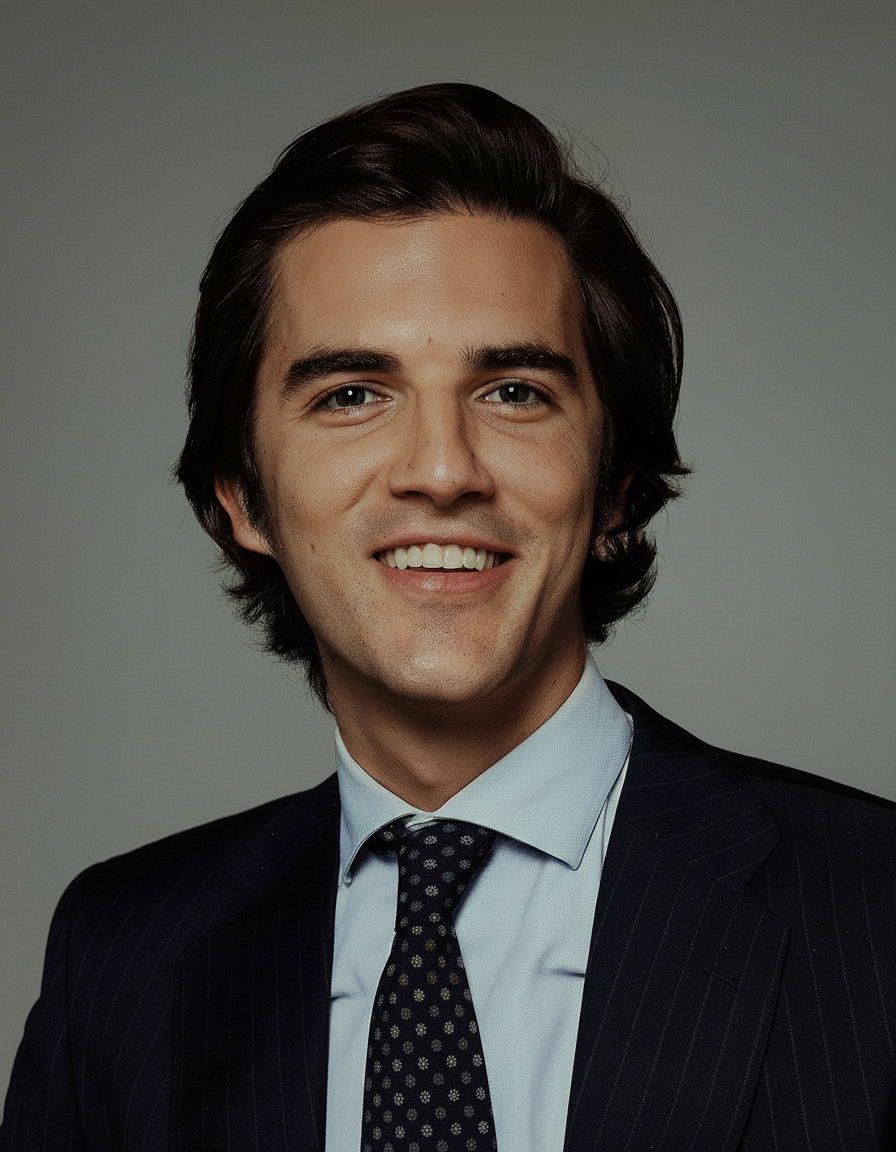
Javier Calatrava Serrano de Haro es odontólogo con dedicación a Cirugía Oral e Implantología, con la periodoncia y cirugía mucogingival como eje de su trabajo. Su actividad clínica se desarrolla en Madrid. Cursó sus estudios de Odontología en Universidad Complutense de Madrid en 2018.
Con el tiempo, ha sumado formación específica en periodoncia y ha llegado al nivel de doctorado. El último de esos programas fue el Doctor en Ciencias Odontológicas, en Universidad Complutense de Madrid (2022).
Su práctica diaria se desarrolla en Clínica Dental Dra. Serrano de Haro, como periodoncista (Madrid, España).
Su aportación al colectivo: imparte 4 formaciones — entre ellas «Straumann Corporate Forum 2026 · Implantología basada en la evidencia» y «Casos complejos en cirugía plástica periodontal, autotrasplante dental guiado y RTG» — en Riyadh Air Metropolitano, Madrid y Guadalajara, México y firma 28 publicaciones científicas en revistas como Clinical and Experimental Dental Research y Clinical Oral Implants Research.
Explora su trayectoria
Doctor en Ciencias Odontológicas
Doctorado · Universidad Complutense de Madrid · Madrid, España
Experto en Clínica Periodontal
Experto · Universidad Complutense de Madrid · Madrid, España
Grado / Licenciado en Odontología
Grado · Universidad Complutense de Madrid · Madrid, España
Clínica Dental Dra. Serrano de Haro · Madrid, España
Sin actividad docente registrada
Enero a abril de 2027 · Módulos I-IV · Madrid - España · Presencial
Odontología Digital · Cirugía Oral e Implantología
22 de octubre de 2026 · Riyadh Air Metropolitano, Madrid · Presencial
Cirugía y Periodoncia · Implantología
27 y 28 de noviembre de 2026 · Guadalajara, México · Presencial
Cirugía Oral e Implantología · Cirugía Plástica Periodontal
Fecha a convenir · Madrid - España · Presencial
Cirugía y Periodoncia
Clinical and Experimental Dental Research
Autores · Łukasz Lassmann, Adam Pióro, Javier Calatrava, H Wang
OBJECTIVES: To present a structured decision-making approach to full-mouth rehabilitation in patients with generalized severe tooth wear, using functional risk classification (Green, Yellow, Red) and the Smile Design and Space (SDS) Concept for vertical dimension planning. MATERIAL AND METHODS: A 65-year-old male presented with generalized erosion and attrition, affecting esthetics and speech. A structured diagnostic protocol began with symptom-based questionnaires and clinical examination. He was categorized as "Yellow" indicating the absence of temporomandibular disorders (TMD), repeatable occlusion, but need for interdisciplinary pretreatment. The SDS Concept guided the vertical dimension of occlusion (VDO) increase, based on incisal display, smile line, and prosthetic space, using the formula A + B - C = VDO. Functional crown lengthening was performed to restore the ferrule. Restorations were done with adhesively bonded lithium disilicate crowns and layered ceramics. RESULTS: A 10 mm VDO increase was well tolerated, with restored esthetics, stable occlusion, and improved phonetics. The SDS protocol provided a reproducible, esthetically driven method for VDO and prosthetic space evaluation with no complications. CONCLUSIONS: Integrating functional risk classification with the SDS concept enables individualized, systematic full-mouth rehabilitation in severe tooth wear cases, improving diagnostic precision and interdisciplinary communication.
Clinical Oral Implants Research
Autores · Muhammad H. A. Saleh, Javier Calatrava, Nataly Rabelo Mina Zambrana, Lucrezia Parma‐Benfenati, Hamoun Sabri, István Urbán, Hom‐Lay Wang
AIMS: To quantify immediate graft displacement following tension-free flap closure in non-contained alveolar ridge defects regenerated using three membrane fixation strategies in an ex vivo human model. METHODS: Eighteen sites from six fresh cadaveric heads with non-contained defects underwent horizontal guided bone regeneration using deproteinized bovine bone mineral and a collagen membrane stabilized by no fixation (NF), periosteal sutures (PS), or titanium fixation pins (FP). Techniques were applied sequentially. Cone-beam CT scans were obtained at baseline, post-grafting, and after passive closure. Linear ridge width was measured at 12 standardized positions, and graft volumes were segmented from STL reconstructions. Mixed-effects multilevel linear regression assessed width changes, while Friedman and paired Wilcoxon tests evaluated volumetric outcomes, with Tukey-adjusted contrasts for intergroup comparisons. RESULTS: ; 44.4% ± 17.9%) (loss p = 0.030; stability p = 0.002). Ridge collapse was greatest with no fixation (-1.37 ± 0.85 mm) compared with PS (-0.40 ± 0.62 mm) and FP (-0.28 ± 0.61 mm). After adjustment, PS and FP reduced collapse by 0.96 mm (p = 0.012) and 1.11 mm (p = 0.004), respectively. CONCLUSION: Even after passive flap release and tension-free closure, membrane fixation, particularly with titanium pins, provides superior resistance to graft displacement in non-contained defects, while unfixed membranes allow substantial graft loss and are therefore not recommended in non-contained defects.
Journal Of Clinical Periodontology
Autores · Yaniv Mayer, Bertha Demetriou, Giulio Rasperini, Eran Gabay, Rok Gašperšič, Darko Božić, Javier Calatrava, Ofir Ginesin, Hadar Zigdon‐Giladi, Ricardo Faría Almeida
AIM: To compare retrieval-augmented systems with general-purpose large language models (LLMs) on standardised periodontal clinical vignettes. MATERIALS AND METHODS: Eleven AI systems were evaluated: nine general-purpose LLMs, one general-purpose retrieval-augmented platform (Perplexity) and one medical-domain retrieval-augmented platform (OpenEvidence). Each responded to 30 synthetic vignettes covering acute, chronic and complex periodontal scenarios. Six blinded periodontists scored responses on a 5-point Likert scale for accuracy, safety, freedom from hallucinations and completeness in a randomised block design. Friedman and Conover-Iman tests with Holm correction were applied; mixed-effects and ordinal models served as sensitivity analyses. RESULTS: At least one parameter scored dangerous (≤ 2) in 3.3%-46.7% of responses across platforms, despite mean composite scores (3.28-4.86) exceeding the rubric midpoint of 3.0. Between-model differences were significant (p < 0.001), with a small-to-medium overall effect (Kendall's W = 0.17) and large within-category effects (W up to 0.82). Perplexity, OpenEvidence and Claude 4.7 Opus formed a top tier. CONCLUSION: Retrieval-augmented systems rated highest, but this advantage was confounded with response length. The dangerous-response spread argues against undifferentiated use. These tools should assist, not replace, specialist judgement.
Clinical Advances in Periodontics
Autores · Kévimy Agossa, D Calzavara, Javier Calatrava, Hom‐Lay Wang
BACKGROUND: The frontal envelope coronally advanced flap (CAF), designed for multiple adjacent gingival recessions (MAGR) of the anterior maxilla, involves tunneling of the midline papilla. However, when treating frontal and lateral MAGR in a single session, the tunneled midline papilla can act as a fixed point, which may limit coronal advancement or lead to mucosal folds in the gingival margin. METHODS: This case report introduces a modification of the CAF technique in which a V-shaped split-thickness surgical papilla is elevated at the midline to enhance flap mobility. Two patients with combined maxillary frontal and bilateral types 1 and 2 (RT1, RT2) MAGR ranging from 2-6 mm were treated using this approach. RESULTS: Postoperative healing was uneventful for both cases. In Case 1, complete and stable root coverage with excellent tissue integration and color match was observed at 18 months. In Case 2 (RT2 MAGR), near-complete root coverage was achieved at 6 months. CONCLUSION: Within the limitations of this case report, the modified CAF appears to be an effective approach for managing MAGR extending beyond the incisors and involving both maxillary quadrants. Clinicians may consider this modification when enhanced coronal flap mobility is required to treat extensive frontal and lateral MAGR in a single session. KEY POINTS: In the traditional frontal envelope coronally advanced flap, the tunneled midline papilla may act as a fixed point, which may restrict coronal advancement or lead to mucosal fold formation in the gingival margin. This case presents a modified design that enhances coronal flap mobility and achieves optimal root coverage in cases of extensive frontal and lateral maxillary anterior gingival recessions (MAGR). The success of this approach relies on midline papilla anatomy and precise incision placement, both of which support adequate flap stability, enhancing the predictability of root coverage outcomes. PLAIN LANGUAGE SUMMARY: Gum recession occurs when the gum margin moves downward, exposing part of the tooth root. When several neighboring teeth in the upper front region are affected, treatment can be particularly challenging because of the esthetic demands in this visible area. A commonly used surgical method, known as the coronally advanced flap (CAF), repositions the gum tissue to cover the exposed roots. However, in its traditional form, the tissue between the two front teeth is not lifted but tunneled, which can limit tissue movement and occasionally cause small folds along the gum line. This report describes a refined version of the CAF technique in which a small, V-shaped flap is gently lifted between the front teeth to improve tissue mobility and adaptation. Two patients were treated using this approach, both achieving favorable healing, stable root coverage, and a natural appearance that blended well with surrounding tissues. This modification may help clinicians manage wider areas of gum recession in the upper front region while maintaining esthetic harmony.
International Journal of Computerized Dentistry
Q3 · IF 1.5Autores · Ignacio Pedrinaci, Amirali Nasseri, Javier Calatrava, Emilio Couso-Queiruga, William V. Giannobile, German O. Gallucci, Mariano Sanz
Coautores Doctor&Cols · Ignacio Pedrinaci Peñalver
AIM: The primary aim of the present in vitro study was to compare methods for generating 3D-printed replicas through virtual segmentation, utilizing artificial intelligence (AI) or manual processes, by assessing accuracy in terms of volumetric and linear discrepancies. The secondary aims were the assessment of time efficiency with both segmentation methods, and the effect of post-processing on 3D-printed replicas. MATERIALS AND METHODS: Thirty teeth were scanned through CBCT, capturing the region of interest from human subjects. DICOM files underwent virtual segmentation through both AI and manual methods. Replicas were fabricated with a stereolithography 3D printer. After surface scanning of pre-processed replicas and extracted teeth, STL files were superimposed to compare linear and volumetric differences using the extracted teeth as the reference. Post-processed replicas were scanned to assess the effect of post-processing on linear and volumetric changes. RESULTS: AI-driven segmentation resulted in statistically significant mean linear and volumetric differences of -0.709 mm (SD 0.491, P 0.001) and -4.70%, respectively. Manual segmentation showed no statistically significant differences in mean linear (-0.463 mm, SD 0.335, P 0.001) and volumetric (-1.20%) measures. Comparing manual and AI-driven segmentations, AI-driven segmentation displayed mean linear and volumetric differences of -0.329 mm (SD 0.566, P = 0.003) and -2.23%, respectively. Additionally, AI segmentation reduced the mean time by 21.8 minutes. When comparing post-processed to pre-processed replicas, there was a volumetric reduction of -4.53% and a mean linear difference of -0.151 mm (SD 0.564, P = 0.042). CONCLUSIONS: Both segmentation methods achieved acceptable accuracy, with manual segmentation slightly more accurate but AI-driven segmentation more time-efficient. Continual improvement in AI offers the potential for increased accuracy, efficiency, and broader application in the future.
Clinical Advances in Periodontics
Q3 · IF 1.2Autores · Ignacio Pedrinaci, Javiera Casas, Samuel Akhondi, Javier Calatrava, David Palombo, Alejandro Lanis, German O. Gallucci
Coautores Doctor&Cols · Ignacio Pedrinaci Peñalver
BACKGROUND: Peri-implant soft tissues are usually reduced when using a tissue punch in flapless static computer-assisted implant surgery (s-CAIS). The purpose of this article is to introduce a technique combining a minimally invasive guided roll flap (GRF) with s-CAIS to enhance buccal mucosal thickness and maintain peri-implant soft tissues. METHODS: A surgical template is used to define a crest-centered de-epithelialization window and to guide a palatal semilunar full-thickness pedicle that is rolled into a mid-buccal tunnel and stabilized. Osteotomy and implant placement are performed through the same template; the transmucosal profile is supported by an immediate provisional restoration, a sealing socket abutment, or a stock healing abutment. Clinical and volumetric outcomes were assessed over a 12-month follow-up. RESULTS: At 12 months, all implants demonstrated increased buccal mucosal thickness and adequate keratinized mucosa width. Volumetric analysis confirmed soft tissue volume augmentation compared to baseline. No vertical loss of adjacent papillae was observed. Clinical parameters, including PPD and BOP, remained stable, indicating maintained peri-implant health. CONCLUSIONS: This technique demonstrates the feasibility of combining s-CAIS with specific surgical approaches to potentially maintain or slightly improve peri-implant soft tissues. It offers clinicians an alternative method to the soft tissue punch, so the peri-implant soft tissues may be maintained or even augmented in a minimally invasive way.
The International Journal of Periodontics & Restorative Dentistry
Q2 · IF 1.9Autores · Ramón Gómez Meda, Javier Calatrava, Jon Misch, Hom-Lay Wang
Bone augmentation procedures often lead to coronal displacement of the mucogingival junction, a deficiency of keratinized mucosa, and a reduction in soft tissue height. These challenges complicate the achievement of an ideal peri-implant phenotype. It is known that addressing both the quantity and quality of soft tissue is crucial for the long-term success and esthetics of implants. This report introduces a novel technique for peri-implant phenotype modification following bone augmentation, which combines a modified onlay graft (MOG) with an apically positioned flap (APF) performed simultaneously with implant placement. Two clinical cases are presented to demonstrate this approach, highlighting the restoration of the soft tissue environment through a single surgical intervention. The technique resulted in successful outcomes, including increased vestibular depth, keratinized tissue width, and vertical soft tissue volume. The MOG technique effectively manages soft tissue in post-bone augmentation scenarios, providing an ideal peri-implant phenotype and leading to stable, esthetic, and functional implant restorations. This method requires fewer surgical interventions than traditional alternatives, minimizing patient discomfort and enhancing overall treatment outcomes.
Journal of Periodontal Research
★ Alto impacto · Q1 · IF 3.4Autores · Agossa K, Sabri H, Chele D, Calatrava J, Bravard M, Wang HL
Aim To assess the effect of connective tissue graft (CTG) in the treatment of periodontal intrabony defects (IDs), focusing on changes in postoperative gingival recession (GR) depth and regenerative outcomes. Methods A systematic search was conducted across MEDLINE–PubMed, EMBASE, Cochrane CENTRAL, and Google Scholar for articles published through December 31, 2024. Randomized controlled trials (RCTs) comparing treatment outcomes in IDs treated with or without CTG were included in a meta‐regression analysis. A mixed‐effect linear regression model was employed to estimate the effect of CTG on postoperative GR depth, probing depth (PPD) reduction, clinical attachment level (CAL) gain, and bone fill (BF). Results Twenty‐three studies were selected, with five RCTs (176 IDs) included in the meta‐regression. Of these, two RCTs compared bone graft (BG) + CTG to BG + resorbable membrane (MB), one compared periosteal vs. nonperiosteal CTG combined with BG, one compared open flap debridement (OFD) + CTG to OFD alone, and one compared BG + CTG to either OFD or MB. The use of CTG was significantly associated with a reduction in GR (mean effect size of 0.981 mm, 95% CI: 0.573 to 1.389, p = 0.001), PPD (mean effect size of 1.160 mm, 95% CI: 0.318 to 2.002, p = 0.010), as well as improvements in CAL (mean effect size of 1.105 mm, 95% CI: 0.420 to 1.790, p = 0.004) and BF (mean effect size of 1.382 mm, 95% CI: 0.595 to 2.169, p = 0.002). Conclusion Within the limitations of the study, the use of CTG in periodontal regenerative therapy for IDs appears beneficial in reducing postoperative GR and might further enhance regenerative outcomes compared to treatments without CTG.
Journal of Oral Implantology
Autores · Jacob Martin Zimmer, Jonathan E. Misch, Paolo Nava, Hamoun Sabri, Javier Calatrava, Hom‐Lay Wang
This article aims to comprehensively address and discuss the thresholds for implant removal in cases of severe peri-implantitis. Electronic databases were searched comprehensively, electronically, and manually. Studies focusing on criteria for explantation of dental implants were included and reviewed comprehensively. Based on the latest evidence, explanation thresholds were proposed. Moreover, the included studies were evaluated and summarized. Current guidelines for implant removal are primarily based on expert opinions rather than actual data. Standardized data encompassing the extent of peri-implant bone loss for assessing treatment outcomes after surgical intervention for peri-implantitis is scarce. Nevertheless, newly available data confirms a poor prognosis for implants with more than 50% bone loss. This supports the recommendation for implant removal in cases of more than 50% bone loss due to peri-implantitis. Implant mobility resulting from the total loss of osseointegration necessitates the removal of the implant. While probing depth provides information about disease severity, it should not be used as the sole diagnostic tool. In the decision-making process for treatment or explantation, factors such as the number of affected implants, the implant's position within the bony housing, intraosseous defect morphology, type of implant restoration, and patient characteristics should be considered. The removal of severely compromised dental implants with peri-implantitis and bone loss exceeding 50% is supported. Moreover, factors related to the restoration, the implant itself, and the patient must be considered when deciding whether to remove or salvage a compromised implant. Clear clinical guidelines for explanting dental implants in cases of severe peri-implantitis need to be included. This poses significant challenges to clinicians when determining whether to explant or treat dental implants compromised by substantial bone loss.
Journal of Periodontology
Autores · Hom‐Lay Wang, Gustavo Ávila‐Ortiz, Alberto Monje, Purnima Kumar, Javier Calatrava, Tara Aghaloo, Shayan Barootchi, Joseph P. Fiorellini, María Elisa Galárraga-Vinueza, Joseph Kan, Guo‐Hao Lin, Andrea Ravidà, Muhammad H. A. Saleh, Lorenzo Tavelli, AO/AAP Consensus Participants, Paul S. Rosen
BACKGROUND: The exponential increase in dental implant use has led to a parallel rise in peri-implant diseases (PID), adversely affecting implant therapy success and patient quality of life. Efforts have been made by the dental community to understand systemic, behavioral, and site-level risk factors involved in the etiologies and pathogenesis of PID and conditions and to develop standardized treatment protocols for the management of these clinical entities. The 2024 Academy of Osseointegration/American Academy of Periodontology (AO/AAP) consensus aimed to integrate the best available evidence and expert opinion into a unified framework for the prevention and management of PID and conditions. METHODS: Focused questions were previously addressed in eight systematic reviews that were grouped into two main topics. Group 1 evaluated systemic and local risk factors/indicators for the development of peri-implant mucositis and peri-implantitis, peri-implant soft tissue deformities, as well as prosthetic factors associated with peri-implant marginal bone loss. Group 2 focused on therapeutic strategies for the management of PID, encompassing nonsurgical debridement, implant surface decontamination methods, and surgical interventions (both nonreconstructive and reconstructive). Structured consensus discussions were held during an on-site meeting in Oak Brook, Illinois (August 14-16, 2024) to inform evidence-based recommendations. RESULTS: A plethora of systemic, behavioral, and local factors may play a pivotal role in the onset and progression of PID and conditions. Key systemic and behavioral risk factors include history of periodontitis, smoking, uncontrolled diabetes, poor microbial biofilm control, and obesity, while implant malposition, unfavorable prosthetic factors, and suboptimal peri-implant soft tissue phenotypical features are relevant site-related factors. Peri-implant mucositis may be effectively managed with nonsurgical debridement and control of risk factors. This possibly represents the first step of treatment of peri-implantitis, whereas more advanced cases require individualized surgical approaches, ranging from flap-for-access, resective, reconstructive, or soft tissue augmentation procedures. Supportive peri-implant maintenance is essential for long-term peri-implant tissue stability and health. CONCLUSIONS: An evidence-based flow diagram combined with expert opinion was generated for clinicians to manage PID and conditions, emphasizing early risk factor identification, tailored treatment protocols, and continued maintenance to optimize long-term implant therapy outcomes.
Journal of Esthetic and Restorative Dentistry
★ Alto impacto · Q1 · IF 5Autores · Ignacio Pedrinaci, Javier Calatrava, Manuel Toledano‐Osorio, Na Zhao, Alejandro Lanis, Mariano Sanz
Coautores Doctor&Cols · Ignacio Pedrinaci Peñalver
Objectives To demonstrate how contemporary digitally driven workflow can enhance outcomes for complex esthetic dental cases, focusing on three distinct clinical scenarios involving implant placement, esthetic crown lengthening, and tooth autotransplantation (ATT). Overview Three multidisciplinary clinical cases demonstrate our contemporary digital workflows, integrating diagnosis, treatment planning, patient communication, and guided execution. The first case involves replacing two anterior central incisors using digital planning, guided surgery, prefabricated customized healing abutments, and a digitally driven restorative process. The second case showcases an esthetic crown‐lengthening procedure, where Multifunctional Anatomical Prototypes (MAPs) serve as both mock‐up and surgical guides, enhancing patient communication on expected outcomes and ensuring precise tissue management to prevent soft tissue rebound. The final case features a tooth ATT, where virtual surgical planning and 3D‐printed tooth replica and guides ensure predictability in the therapeutic outcome. These digitally enabled strategies underscore the predictability and customization achievable with contemporary dental technology. Conclusions Dental treatments in the esthetic zone require meticulous planning and precise execution to achieve controlled results that ensure patient satisfaction and long‐term stability. Contemporary Digital Dentistry enhances predictability, creating a pathway that leads to success.
The International Journal of Oral & Maxillofacial Implants
Autores · Mingfu Ye, Wenjun Liu, Javier Calatrava, Hom‐Lay Wang, Wenxia Huang
PURPOSE: To examine whether mandibular overdentures supported by a single implant vs two implants have similar clinical outcomes, especially pertaining to implant survival, marginal bone loss (MBL), and prosthetic complications. MATERIALS AND METHODS: A systematic review and meta-analysis of randomized controlled trials (RCTs) was conducted. Scientific databases (PubMed, Embase, Scopus, Web of Science, and Cochrane Library) were searched from inception until October 2024. These databases were searched for RCTs comparing single- implant- and two- implant-supported mandibular overdentures that reported implant failures, MBL, and prosthetic complications. RESULTS: Eleven RCTs were included. The meta-analysis showed reduced implant failure rates with single implants at 1 year (OR: 0.33; 95% CI: 0.10, 1.15; I2 = 0%) and 5 years (OR: 0.11; 95% CI: 0.01, 2.16), but the results were not statistically significant. However, pooled subgroup analyses of studies with a follow-up period of 2 to 3 years indicated a significantly reduced implant failure rate with a single implant compared to two implants (OR: 0.12; 95% CI: 0.03, 0.54; I2 = 0%). No significant differences between the implant failure rates were observed between the two groups based on implant loading protocols. Meta-analysis also showed that there were no significant differences in the risk of MBL between single- and two-implant groups (MD: -0.15; 95% CI: -0.31, 0.01; I2 = 43%). The pooled analysis also showed that the risk of overdenture fracture, relining, and metal housing reattachment did not differ significantly between single- and two-implant groups. CONCLUSIONS: Evidence from a limited number of RCTs, mainly with small sample sizes, indicates that single- implant-supported mandibular overdentures may have a tendency to cause implant failures at a lower rate than two- implant-supported mandibular overdentures. The risk of MBL and prosthetic complications may not differ between the treatment modalities.
Journal of Clinical Periodontology
★ Alto impacto · Q1 · IF 5.061Autores · Sabri H, Hazrati P, Tavelli L, Garaicoa-Pazmino C, Calatrava J, Wang HL, Barootchi S
Objective To investigate the associations between peri‐implant phenotype characteristics and long‐term outcomes of soft tissue–level implants. Methods Twenty‐five tissue‐level implants from a previous controlled clinical trial were evaluated at 1‐ and 5‐year follow‐ups after crown delivery. Data included ultrasonographic scans (mucosal thickness and supracrestal tissue height), standardised 2D radiographs, cone beam computed tomography, clinical outcomes (mucosal recession, probing pocket depth, bleeding on probing), patient‐reported outcomes and peri‐implant health status. Standard logistic and linear regression models were used to analyse associations of implant‐ and patient‐related factors with outcomes, including peri‐implant disease status, mucosal recession and marginal bone level (MBL) changes. Results Five‐year implant survival was 100%, with peri‐implant mucositis diagnosed in 36% of the implants. Mean MBL changes from the first to the fifth year was 0.29 ± 0.29 mm. Baseline (pre‐surgical) buccal soft‐tissue thickness < 1.5 mm (OR: 5.20, p = 0.007) and increased plaque scores (OR: 30.11, p < 0.001) were associated with peri‐implant mucositis, whereas buccal bone thickness ≥ 1.5 mm (OR: 0.48, p = 0.039), mucosal thickness around implant (OR: 0.20, p = 0.001) and supracrestal tissue height (OR: 0.50, p = 0.002) emerged as protective factors. Mucosal recession was significantly associated with baseline buccal soft‐tissue thickness < 1.5 mm (estimate = 0.27, p = 0.03) and keratinised mucosa width of < 2 mm (estimate = 0.39, p = 0.021). Clinical thresholds associated with long‐term peri‐implant health were defined as follows: supracrestal tissue height ≥ 2.8 mm, restorative emergence angle ≤ 35.5°, mucosal thickness 1.8 mm and buccal bone thickness 1.5 mm. Patients reported a high overall satisfaction (visual analogue scale: 88.2%). Colour Doppler ultrasonography showed a strong correlation between tissue perfusion and peri‐implant disease ( r = 0.93, p < 0.001). Conclusions Within the limitations of this study, tissue‐level implants showed excellent survival rates and patient satisfaction at 5 years. Several peri‐implant parameters—such as supracrestal tissue height ≥ 2.8 mm, restorative emergence angle ≤ 35.5° and buccal bone thickness ≥ 1.5 mm—were associated with favourable outcomes. These exploratory thresholds may be considered hypothesis‐generating and could help guide future research and clinical monitoring, although validation in larger cohorts is essential. Trial Registration: www.clinicaltrials.gov/study/NCT02925078
Periodontology 2000
★ Alto impacto · Q1 · IF 8Autores · Zhaozhao Chen, Lan-Lin Chiou, Javier Calatrava, Hom-Lay Wang
Antibiotics are commonly prescribed in periodontal and implant surgeries, either before, during or after surgery, to prevent postoperative infection and reduce early implant failure. However, the potential benefits may be undermined by the risks of resistance and sensitization, thus requiring a strict indication for prescription. There are controversial views regarding the indication, dosage, duration, and timing of antibiotic medication in periodontal and implant surgery. Therefore, the aim of this review is to address the benefits and concerns regarding the efficacy of using preventive antibiotic therapy in Periodontology and Implant Dentistry. Specifically, various types of procedures were comprehensively explored, and recommendations were given accordingly. The available evidence indicates that preventive antibiotic therapy is not warranted for periodontal surgeries, including conventional, plastic, and regenerative procedures. While antibiotic prophylaxis has shown effectiveness in preventing early implant failures in straightforward implant placements, postoperative coverage does not appear to be justified. Furthermore, antibiotic administration has not proven to significantly reduce early implant failure rates in implant placements involving simultaneous guided bone regeneration. There remains insufficient evidence to support or oppose the use of preventive antibiotic therapy for reducing infection rates after ridge or sinus augmentation procedures, nor is there evidence favoring any specific protocol over others.
Journal Of Clinical Periodontology
Autores · Hamoun Sabri, Paolo Nava, Parham Hazrati, Abdusalam Alrmali, Pablo Galindo‐Fernández, Muhammad H. A. Saleh, Javier Calatrava, Shayan Barootchi, Lorenzo Tavelli, Hom‐Lay Wang
AIM: To assess the reliability of ultrasonographic, cone beam computed tomographic (CBCT), probe transparency and transgingival probing (TGP) methods in evaluating gingival thickness (GT), compared with the gold standard histological assessment. METHODS: Sixteen fresh cadaver heads with intact gingivae were used. The sequence for GT measurement included CBCT, ultrasonography, probe transparency, TGP and histology. Both stainless steel periodontal probe and colour-coded probes were used for transparency. TGP involved a calibrated endodontic spreader, and histologic samples served as a comparative standard. Primary outcomes evaluated accuracy in GT measurement, while secondary outcomes assessed agreement among methods and established an optimal threshold for thin versus thick gingiva. RESULTS: One hundred and fifteen teeth were examined, yielding a mean GT of 1.34 mm histologically. US and CBCT underestimated GT (means of 1.25 mm and 1.13 mm, respectively), while TGP overestimated (1.51 mm). Correlations (r = 0.88-0.98) and ICC values (0.73-0.95) indicated strong inter-method agreement. Regression models significantly estimated histological GT from US, CBCT and TGP. A new 1.18 mm cut-off, based on histology, improved diagnostic accuracy over the traditional 1 mm threshold. CONCLUSIONS: While histology remains the GT reference standard, US, CBCT and TGP achieved clinically acceptable accuracy. US showed the highest agreement with histology, followed by TGP and CBCT. The study supports US as the most practical non-invasive tool, although CBCT and TGP remain viable options. Further clinical validation is recommended, acknowledging the limitations of cadaveric models in reflecting in vivo conditions.
Periodontology 2000
Autores · Hom‐Lay Wang, Parham Hazrati, Javier Calatrava, Muhammad H. A. Saleh, Abdusalam Alrmali
This systematic review and meta-analysis aimed to evaluate the long-term clinical outcomes of regenerative procedures compared with access flap surgery for the treatment of intrabony defects, with a minimum follow-up period of 5 years. A systematic review protocol following PRISMA guidelines was conducted. Both electronic and manual searches were conducted to identify randomized clinical trials (RCTs) on regenerative treatment of deep intrabony defects (≥3 mm) with a follow-up of at least 5 years. Primary outcome variables were probing depth (PD) reduction, clinical attachment level (CAL) gain, recession depth (REC) and tooth loss. Meta-analyses and meta-regressions were performed using random-effects models. Seventeen RCTs published from 2004 to 2022, accounting for 501 defects, with follow-ups ranging from 5 to 20 years, were included. Thirteen studies with some concerns and four with high risks of bias were identified. Meta-analyses revealed that after ≥5 years of follow-up, guided tissue regeneration (GTR) on the intrabony defect resulted in significant CAL gain (3.27 mm; 95% CI: 2.90-3.65) and PD reduction (4.04 mm; 95% CI: 3.69-4.38) compared with baseline. After ≥5 years, regenerative procedures with biologics, bone grafts, or both showed significant improvements in CAL gain (3.21 mm; 95% CI: 2.72-3.70) and PD reduction (3.92 mm; 95% CI: 3.39-4.44). GTR on the intrabony defects obtained higher long-term CAL gain (1.52 mm; 95% CI: 0.06-3.10) and PD reduction (0.89 mm; 95% CI: 0.22-1.99) than OFD (open flap debridement); however, none of the outcomes reached statistical significance (p = 0.06; p = 0.115). Meta-regression identified significant associations between outcomes and factors, such as follow-up time, surgical technique, membrane type, and baseline measurements. The certainty of evidence was low for CAL and PD outcomes, but high for REC. Long-term studies indicate that regenerative procedures for the intrabony defects, particularly GTR, provide significant improvements in clinical parameters compared with baseline. However, the evidence does not conclusively demonstrate the superiority of regenerative approaches over OFD in the long term.
Clinical Advances in Periodontics
Autores · Mang Ye, Dumitru Chele, Javier Calatrava, Abdusalam Alrmali, Wenxia Huang, Hom‐Lay Wang
BACKGROUND: The ultimate objective of implant dentistry is to position the implant in a three-dimensional, prosthetic-driven location. This case highlights a guided approach for harvesting and positioning an autogenous bone block to restore a horizontal bone defect in the anterior maxilla. METHODS: This case report describes a 55-year-old patient with horizontal bone deficiency in the anterior maxilla following teeth loss. Using specialized software, a surgical guide was designed to facilitate the harvesting of an autogenous bone block from the subnasal region in the same location where implants were planned to be placed. The graft was then repositioned and fixed with titanium screws, and the gaps were grafted with xenogenic bone particles and covered with an absorbable collagen membrane. After 6 months, the implants were placed, followed by prosthetic restoration. RESULTS: A one-stage implant placement was performed after an uneventful healing period. The bone augmentation resulted in a ridge width of 8 mm for a net gain of 5 mm. After 4 months, the implants were loaded with a screw-retained zirconia bridge. CONCLUSION: Guided transpositional bone blocks offer a predictable approach to treating horizontal bone defects in the esthetic zone. Utilizing digital planning and surgical guides enhances precision, making the result more predictable. KEY POINTS: This case provides new information as it highlights a novel guided approach for harvesting and positioning an autogenous bone block to restore a horizontal bone defect in the anterior maxilla using a surgical guide. The keys to successful management of this case include using precise digital planning, the design and use of a surgical guide to accurately harvest the autogenous bone block, proper fixation of the graft and ensuring an uneventful healing period before implant placement and prosthetic restoration. The primary limitations to success in this case could involve the challenge of having adequate distance away from the nasal floor for harvesting and repositioning the autogenous bone block and potential complications during the healing period. PLAIN LANUAGE SUMMARY: This report describes a modern technique for addressing bone loss in the upper front part of the mouth, crucial for placing dental implants correctly. A 55-year-old patient with insufficient bone was treated using a digital plan to precisely guide the movement of a bone piece from a nearby area to where it was needed. This guided approach involved designing a custom guide with computer software, securely attaching the bone, and using special materials to aid healing. After 6 months, the dental implants were successfully placed and fitted with new teeth, resulting in a stable and natural-looking outcome.
BMC Oral Health
Autores · Mingfu Ye, Xiuwen Lin, Wenjun Liu, Javier Calatrava, Wenxia Huang, Hom‐Lay Wang
PURPOSE: Perforation of the sinus membrane is a common complication during maxillary sinus lift surgery. This systematic review aimed to evaluate if the residual ridge height (RRH) is a risk factor for membrane perforations during the lateral-window sinus lift surgery. METHODS: PubMed, Embase, CENTRAL, Scopus, and Web of Science databases were searched for studies reporting the association between RRH and sinus membrane perforations during the lateral-window sinus lift surgery. The last date of the search was 17th June 2024. RESULTS: Ten studies were eligible. 1601 patients undergoing 1809 sinus lift procedures were included in the studies. The overall sinus membrane perforation rate was 19.2%. The meta-analysis found that mean RRH was significantly lower in the perforation group than the non-perforation group (MD: -0.89 95% CI: -1.47, -0.31 I2 = 90%). The included studies classified Small RRH as a 3–4 mm ridge. Meta-analysis showed a tendency of increased risk of perforation with small RRH, but the difference was not statistically significant (OR: 2.47 95% CI: 0.49, 12.21 I2 = 88%). The exclusion of one outlier study showed statistically significant results. CONCLUSIONS: Small RRH seems to be a potential risk factor for sinus membrane perforation during lateral-window sinus lift surgery. More evidence is needed for improving the validity of the current results.
Clinical Oral Implants Research
★ Alto impacto · Q1 · IF 4.3Autores · Ignacio Sanz-Sánchez, Ana Molina, Conchita Martin, Juan Bollain, Javier Calatrava, Mariano Sanz
OBJECTIVE: The aim of this study was to evaluate the long-term (5 years) clinical efficacy of the one-abutment one-time protocol (test) versus the standard of care by placing the definitive abutment on the day of the prosthetic delivery (control). MATERIALS AND METHODS: In this study, 39 subjects with 60 implants were randomly allocated to either the test or the control group. Changes in the radiographic interproximal bone levels (DIB), modified sulcus bleeding index, probing depth, modified plaque index, papilla fill (Jemt score), incidence of peri-implantitis and peri-implant mucositis as well as patient-reported outcomes measures (PROMs) were collected and compared at 1, 3 and 5 years. RESULTS: At 5 years, the control group showed a greater, although not statistically significant, change in mean DIB values (0.97 mm vs. 0.53 mm). Regarding the other clinical parameters evaluated, no statistically significant differences were observed between groups at any time point. At 5 years, 51% of the implants presented peri-implant mucositis (25.5% in the control and 23.5% in the test), and only one implant in the test group developed peri-implantitis. CONCLUSIONS: The connection and disconnection of healing abutments during the healing period was not associated with higher long-term bone loss. Clinical outcomes and PROMs were similar between groups.
Clinical Implant Dentistry and Related Research
Autores · Paolo Nava, Hamoun Sabri, Javier Calatrava, Jacob Martin Zimmer, Zhaozhao Chen, Junying Li, Hom‐Lay Wang
OBJECTIVE: To evaluate the feasibility of ultrasound-image-based computer-assisted implant planning and placement. MATERIALS AND METHODS: Intraoral scans, cone-beam computerized tomography (CBCT), and ultrasound (US) scans with a custom positioning device were acquired in nine patients. Prosthetic-driven surgical guides were planned and fabricated based on ultrasound images and intraoral scans. Implants were then placed. Postoperative implant position was obtained intra-surgically by intraoral scan. Aside from the ultrasound-based plan, conventional implant planning was performed by the same operator on a pre-surgical CBCT for comparison. Linear deviations between ultrasound and CBCT-planned implant positions were measured and compared with the intra-surgical implant position, and the position deviations between two consecutive plannings were performed on the same CBCT by the same operator. The linear deviation between the 3D scan surface of the edentulous region and the ultrasonographic soft tissue profile segmentation was also assessed with reverse-engineering software. Means, standard deviations, and root mean square differences (RMSD) were calculated for every variable. RESULTS: All the ultrasound-planned implants were successfully placed, and no complications were recorded. The mean deviations in angles, shoulders, and apexes were 5.27 ± 1.75° (RMSD: 5.53°), 0.92 ± 0.26 mm (RMSD: 0.95 mm), and 1.41 ± 0.61 mm (RMSD: 1.53 mm), respectively, between the US and CBCT-planned implants; 2.63 ± 0.43° (RMSD: 2.66°), 1.16 ± 0.30 mm (RMSD: 1.19 mm), and 1.26 ± 0.27 mm (RMSD: 1.28 mm) between the planned implant and intra-surgically recorded positions; and 2.90 ± 1.36° (RMSD: 3.18°), 0.65 ± 0.27 mm (RMSD: 0.70 mm), and 0.99 ± 0.37 mm (RMSD: 1.05 mm) between two consecutive CBCTs planning performed by the same operator. The mean deviation between the 3D surfaces of model scans and ultrasound-derived soft tissue profile in the edentulous area was 0.19 ± 0.08 mm. CONCLUSIONS: Ultrasound-guided implant surgery represents a feasible non-ionizing alternative to conventional static guided implant surgical protocols for implant placement in sites with favorable characteristics.
Clinical Implant Dentistry and Related Research
★ Alto impacto · Q1 · IF 3.598Autores · Junying Li, Zhaozhao Chen, Paolo Nava, Shengtao Yang, Javier Calatrava, Hom-Lay Wang
Objective: To assess a newly developed intraoral scan protocol in enhancing the accuracy of complete-arch implant impressions. Materials and Methods: Four impression approaches were applied to the same maxillary edentulous model with 6 implants: (1) intraoral scan (IOS), (2) intraoral scan with scan aid (IOS-SA), (3) calibrated intraoral scan protocol (CISP), and (4) conventional splinted open-tray impression (CONV). Each approach was repeated 10 times, and a direct scan of the model with a desktop scanner was used as a reference model. The alignment of scans and the reference model was conducted by two methods: (a) aligning all scan bodies to evaluate the overall fit, and (b) aligning the first and second scan bodies to simulate the Sheffield fit test for passive fitting of multiple implant-supported prostheses. Linear deviations from the reference model (trueness) and within each group (precision) were analyzed using Python scripts. Results: When aligned by all scan bodies, the CISP group exhibited comparable mean trueness (38.33 μm) and precision (45.97 μm) to the CONV group (44.30 and 47.92 μm respectively), both of which significantly outperformed the IOS group (86.82 and 83.17 μm respectively). Furthermore, in the virtual Sheffield fit test, the CISP group achieved the highest levels of mean trueness at the end span (121.7 μm), making a linear deviation reduction of 36.7%, 60%, and 41.4% when compared to the CONV, the IOS, and the IOS-SA groups, respectively. Moreover, the CISP group (104.3 μm) displayed a remarkable 65, 182, and 86 μm advantage in precision over the CONV, IOS, and IOS-SA groups, respectively. Conclusion: CISP demonstrated comparable accuracy to the gold standard, the conventional splinted open-tray impression. Furthermore, it excelled in the virtual passive fitting test.
Clinical Implant Dentistry and Related Research
Autores · Javier Calatrava, Ignacio Sanz‐Sánchez, Ana Molina, Juan Bollaín, Conchita Martín, Mariano Sanz
OBJECTIVE: The aim of this investigation was to evaluate the effect on clinical and radiological outcomes of the one-abutment, one-time protocol (test) versus placing the definitive abutment on the day of functional loading after having disconnected and connected three times the healing abutment during the prosthetic phase (control). MATERIALS AND METHODS: Forty patients with 80 implants were randomly allocated to either the test or the control group. Changes in the radiographic marginal bone levels (MBLs), clinical outcomes, prosthetic-related outcomes, and patient-reported outcomes measures (PROMs) were assessed and compared 6 and 12 months after functional loading. RESULTS: Thirty-seven patients with 74 implants were followed at 12 months. A statistically significant bone remodeling was observed in both groups following implant placement. MBLs were significantly greater in the control group at the 6- (-0.13 mm vs. -0.61 mm) and 12-month visits (-0.01 mm vs. -0.53 mm). Bone loss was significantly greater in the control group from surgery to 6 and 12 months and from loading to 6 and 12 months. The abutment height was significantly greater in the test group, however, there were no significant differences in the restorative angle. Similarly, there were no statistically significant differences between groups for the measured clinical variables (probing depth, plaque, and bleeding index) and PROMs. CONCLUSIONS: Disconnecting and reconnecting the healing abutment was associated with significantly higher bone loss after 12 months, as compared to the placement of the definitive abutment at implant installation.
The International Journal of Periodontics & Restorative Dentistry
Autores · P Khayat, David Aidan, Javier Calatrava, Hom‐Lay Wang
Implantoplasty involves mechanically modifying the exposed implant surface by removing macro- and microstructures and is typically considered a resective approach. The potential for bone regen-eration with implantoplasty alone has not yet been studied. This research aimed to evaluate long-term changes in peri-implant bone levels without using regenerative materials. A total of 56 patients (70 implants) were included in the study and were followed for up to 10 years, with a mean observa-tion time of 59 months (~5 years). The implants were evaluated retrospectively through radiograph-ic and clinical (bleeding on probing and suppuration) analyses. Of the 70 implants, 11 (15.7%) were lost due to disease recurrence and additional bone loss. One implant (1.4%) experienced up to 1.0 mm of bone loss, 15 implants (21.4%) showed no change in marginal bone levels (MBLs), and 43 implants (61.5%) gained bone up to 4.8 mm, all without the use of any graft material or membrane. The overall mean MBL change for the remaining 59 implants at the end of the follow-up period (84.3%) was a gain of 1.27 mm. Implantoplasty alone without osseous surgery, flap repositioning, or use of a graft material or membrane may be a viable option for managing peri-implantitis defects. However, future randomized clinical trials with larger sample sizes are necessary to validate current findings.
International Journal of Oral Implantology (Berlin, Germany)
★ Alto impacto · Q1Autores · Hamoun Sabri, Teresa Heck, Neshatafarin Manouchehri, Sara Alhachache, Javier Calatrava, Craig M. Misch, Hom-Lay Wang
PURPOSE: To review and compare the available literature on bone regeneration using titanium mesh and map the current evidence on bone gain outcomes and complications while comparing this scaffold with collagen membranes. MATERIALS AND METHODS: A comprehensive electronic and manual search was performed to identify randomised and non-randomised prospective controlled clinical trials that involved the use of titanium mesh in at least one arm, with outcomes including complications and vertical and/or horizontal bone gain. The focused questions were defined as follows: What are the outcomes of using titanium mesh in ridge augmentation compared to other types of barrier membrane, and what is the complication rate (membrane exposure and infection) when titanium mesh is used in these procedures? RESULTS: A total of 22 articles were included in the qualitative analysis. Overall, the studies that measured bone gain resulted in 3.36 mm vertical (196 subjects; 95% confidence interval 2.44 to 4.64 mm, range 1.4 to 5.7 mm) and 3.26 mm horizontal augmentation (81 subjects; 95% confidence interval 2.93 to 3.63 mm, range 2.6 to 3.7 mm), with variability among studies. The most commonly noted complication was mesh exposure, regardless of the type of mesh used, and the second most common was graft failure. The overall pooled complications rate reported in clinical trials was 10.8%. The meta-analysis comparing titanium mesh and collagen membranes, controlling for the type of bone regeneration (staged or simultaneous with implant placement), failed to show a significant difference in horizontal bone gain between the two techniques. CONCLUSIONS: Within the limitations of the present study and acknowledging the heterogeneity among the articles included, titanium mesh can serve as a feasible protective scaffold for bone regeneration with a relatively acceptable complication rate and in defects requiring around 4 mm 3D reconstruction. Data on patient-reported outcomes were scarce.
Journal of Dentistry
★ Alto impacto · Q1 · IF 4.94Autores · Ignacio Pedrinaci, Javier Calatrava, Emilio Couso-Queiruga, Juan del Rosal Bethencourt, Ignacio Sanz-Sanchez, German O. Gallucci, Mariano Sanz
Coautores Doctor&Cols · Ignacio Pedrinaci Peñalver
OBJECTIVES: Digital protocols and bioactive materials may reduce complications and improve tooth autotransplantation (ATT) success and survival rates. This prospective study assesses the performance of a fully digital autotransplantation protocol of close-apex molars with the adjunctive application of Enamel Matrix Derivatives (EMD). METHODS: Twelve adult patients with 13 hopeless molar teeth were replaced with autotransplantation of closed apex third molars. Outcomes, including success and survival rates, clinical, endodontic, radiographic, patient-reported outcome measures (PROMs), and digital image assessments, were conducted over a two-year follow-up period. RESULTS: Survival and success rates were 100% and 91.2%, respectively, with no progressive inflammatory or replacement root resorption (ankylosis) except for one tooth presenting radiographic furcation involvement. A significant probing depth reduction of 2.4 ± 2.58 mm and CAL gains of 2.8 ± 3.03 mm were observed in transplanted teeth compared to the hopeless receptor teeth. Radiographic bone levels remained stable throughout the study period (-0.37 ± 0.66 mm), and digital image assessments showed minimal alveolar ridge width changes (-0.32 to -0.7 mm) and gingival margin changes (-0.95 to -1.27 mm) from baseline to last visit. PROMs indicated very high patient satisfaction. CONCLUSION: The use of a digital ATT protocol with adjunctive use of EMD in closed-apex third molars demonstrated promising short-term high success and survival rates. Additionally, this type of therapy adequately preserves the dimensions of the alveolar ridge in the receptor site. CLINICAL SIGNIFICANCE: This is the first prospective clinical study examining the effect of a digital tooth autotransplantation protocol combined with the application of EMD. It demonstrates that this approach is an effective treatment for replacing hopeless teeth and also validates the digital assessment of ATT alveolar ridge preservation at the recipient site.
The International Journal of Periodontics & Restorative Dentistry
Autores · Tiziano Testori, Massimo Simion, F. Giachi Carù, Stefano Corbella, Silvio Taschieri, G. Perrotti, Renato Cocconi, Javier Calatrava, Ann M. Decker, Hom‐Lay Wang
Tooth autotransplantation is a reliable procedure with high long-term success and survival rates and shows many advantages over dental implants, particularly for young patients in craniofacial devel-opment. Nonetheless, insufficient bone availability at the recipient site is a key limitation. This study aimed to establish the feasibility of tooth autotransplantation combined with staged or simultaneous bone regeneration to address complex cases. Two young patients with missing teeth due to hypodon-tia or trauma and who were deemed unsuitable for implants were treated. Both complex cases were addressed through autologous tooth autotransplantation, either in conjunction with simultaneous guided bone regeneration or into a previously regenerated crest. The 5-year follow-up for the auto-transplanted canine and more than 20-year follow-up for the autotransplanted premolar revealed satisfactory results. These teeth played pivotal roles in restoring both function and esthetics in young patients. Additionally, tooth vitality was preserved throughout the follow-up period, with no compli-cations noted. Tooth autotransplantation in combination with previous or simultaneous guided bone regeneration proved to be effective in restoring function and esthetics in complex situations in young patients.
Journal of Esthetic and Restorative Dentistry
★ Alto impacto · Q1 · IF 5Autores · Ignacio Pedrinaci, Javier Calatrava, Juan Flores, Adam Hamilton, German O. Gallucci, Mariano Sanz
Coautores Doctor&Cols · Ignacio Pedrinaci Peñalver
Objective To describe a strategy using digital technologies for improving the diagnosis, treatment planning, and surgical execution of patients with excessive gingival display (EGD) due to altered passive eruption (APE). Clinical Considerations An important component for successful patient's management is to fulfill their esthetic expectations whilst delivering predictable and long‐term therapeutic outcomes. To achieve this goal in patients with excessive gingival display due to altered passive eruption, it is essential to perform an accurate diagnosis and to communicate to the patient the expected customized results using digital technologies. Computer‐aided designed and manufactured multifunctional anatomical prototypes (MAPs) may contribute to these purposes. Additionally, they can guide the surgical crown lengthening procedure or serve as a reference during the surgical guide fabrication providing information of the required anatomical landmarks. Conclusions This novel strategy protocol for diagnosis, communication, and treatment management of patients with excessive gingival display follows functional and biological principles within the frame of a digital workflow, which improves the diagnostic capabilities, enhances communication, and guides the surgical treatment as shown in the 12 months follow‐up of the reported case. Clinical Significance Developing a virtual patient by combining multiple digital data sets including cone‐beam computed tomography (CBCT), intra‐oral scans and digital photography, supports the clinician and the patient to achieve a comprehensive diagnosis and to better communicate the expected results to the patient. Furthermore, this digital treatment exercise based on anatomical and biological principles will facilitate the surgical precision and the achievement of successful outcomes, thus fulfilling the patient needs and expectations.
Periodoncia Clínica (SEPA) nº23 · Monográfico 'Science and practice of tooth autotransplantation'
Autores · Pedrinaci I, Calatrava J, Sanz-Sánchez I, Sanz M
Coautores Doctor&Cols · Ignacio Pedrinaci Peñalver
Los autotrasplantes dentales presentan tasas de supervivencia superiores al 90% con seguimientos de más de 20 años, y su éxito depende directamente de la vitalidad del ligamento periodontal del diente donante. Se presenta el caso de un varón de 17 años con un primer molar inferior izquierdo no restaurable, sustituido por el tercer molar del mismo cuadrante mediante un flujo de trabajo completamente digital: CBCT convertido a STL, modelo estereolitográfico mandibular impreso en 3D, réplica del diente donante y férula de cirugía guiada para la osteotomía del alvéolo receptor. Antes de reposicionar el diente se aplicaron proteínas derivadas de la matriz del esmalte (EMD) sobre la superficie radicular para aumentar el potencial regenerativo y reducir las complicaciones del procedimiento.
Cirugía Oral e Implantología
Islas Baleares · España
Cirugía Oral e Implantología
Valencia · España
Cirugía Oral e Implantología
Málaga · España
Cirugía Oral e Implantología
Sevilla · España
Cirugía Oral e Implantología
Madrid · España
Cirugía Oral e Implantología
Alicante · España