Endodoncia y Cirugía Apical
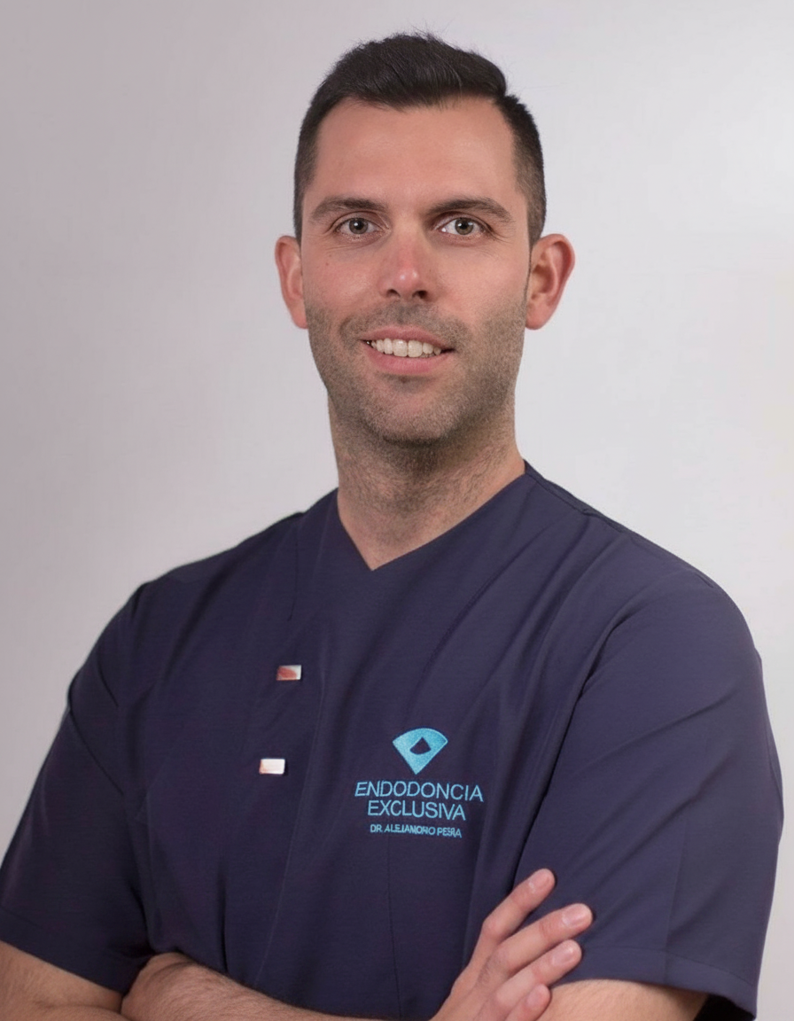
Alejandro Peña López es odontólogo con dedicación a Endodoncia y Cirugía Apical. Su actividad clínica se desarrolla en Almería. Su formación de base procede de Universidad Europea en 2011.
A lo largo de su trayectoria, ha llegado al nivel de doctorado. El último de esos programas fue el Doctorado en Odontología, en Universidad Católica San Antonio de Murcia (UCAM) (2017). Ha pasado por Universidad Católica San Antonio de Murcia (UCAM) y Universidad Europea.
Actualmente ejerce como endodoncista en Clínica dental Altamira, Centro dental DEA y Clínica dental Rovira en Madrid y Almería. Previamente pasó por Clínica dental Dr. Giménez y Clínica dental Frasdent.
La docencia ocupa la otra mitad de su tiempo: imparte clase en Universidad Católica San Antonio de Murcia (UCAM), en programas como Máster en Endodoncia Clínica y Microscópica.
En Doctor&Cols imparte una formación — entre ellas «Curso Intensivo de Actualización en Endodoncia · Sevilla 2026» — en Sevilla, España, ha publicado 3 casos clínicos sobre endodoncia en conductos calcificados, endodoncia y ultrasonidos y firma 3 publicaciones científicas en revistas como The Journal of the American Dental Association y Australian Endodontic Journal.
Explora su trayectoria
Doctorado en Odontología
Doctorado · Universidad Católica San Antonio de Murcia (UCAM) · Murcia, España
Curso de técnicas microscópicas y piezoeléctricas en odontología
Otro · Centro de Cirugía de Mínima Invasión Jesús Usón · Cáceres, España
Máster Universitario en Endodoncia Avanzada
Máster · Universidad Europea · Madrid, España
Programa de Capacitación Clínica
Otro · Universidad Europea · Madrid, España
Grado / Licenciado en Odontología
Grado · Universidad Europea · Madrid, España
Clínica dental Altamira · Madrid, España
Centro dental DEA · Almería, España
Clínica dental Rovira · Almería, España
Clínica dental Dr. Giménez · Madrid, España
Clínica dental Frasdent · Madrid, España
Universidad Europea (Clínica Universitaria) · Madrid, España
Coordinador · Máster en Endodoncia Clínica y Microscópica
Universidad Católica San Antonio de Murcia (UCAM) · Murcia, España
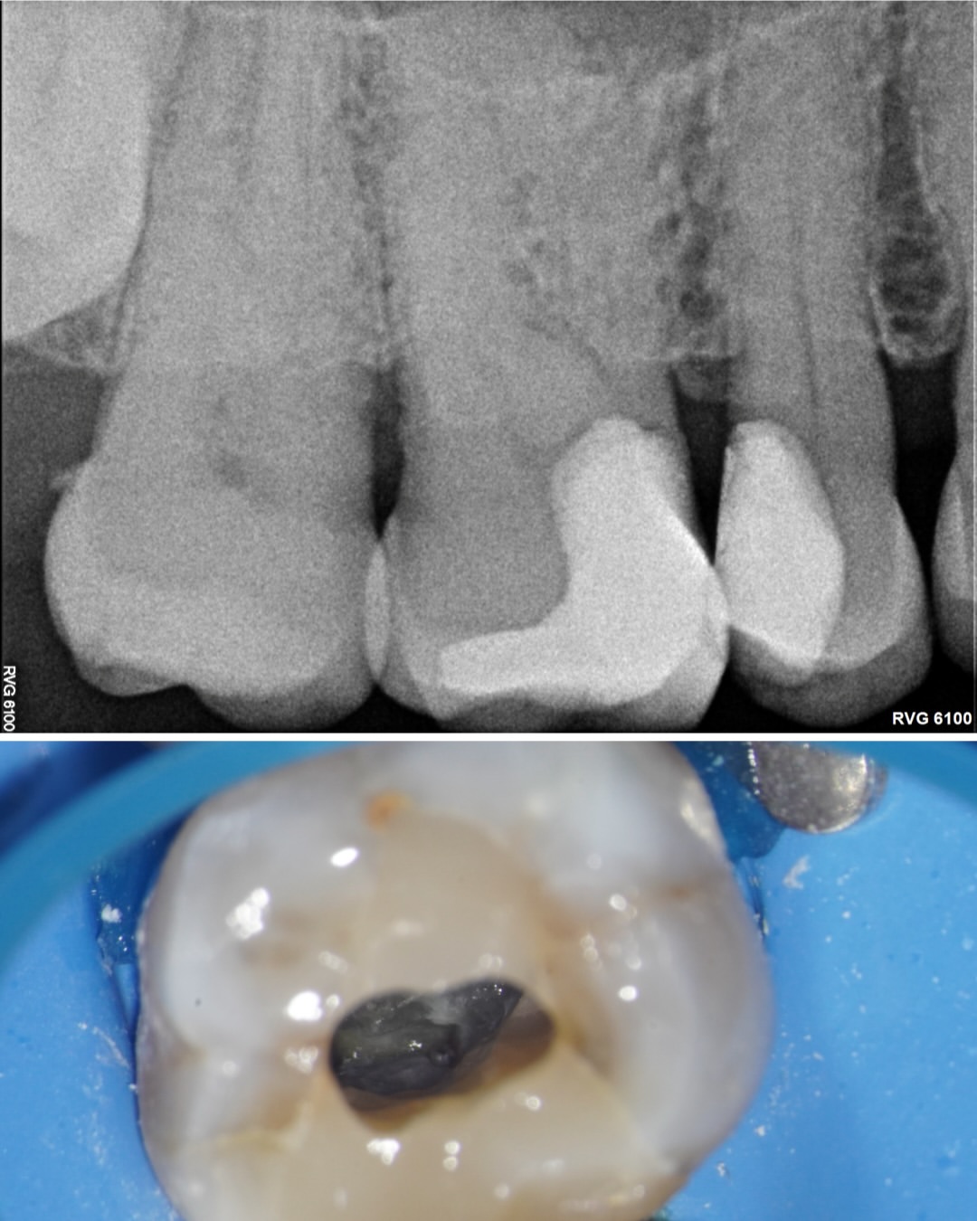 5 imgs
Manejo de una curvatura de 90° en la raíz distovestibular de un 16 con metamorfosis cálcica: ultrasonidos para localizar los conductos, limas manuales 06 y 08 para negociar la curvatura distal y glidepath con RaceEvo 02.
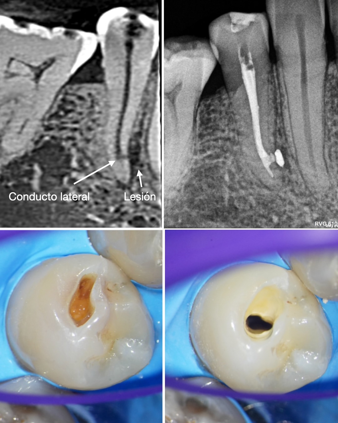
Un segundo premolar inferior con caries y lesión laterorradicular resuelto con apertura conservadora a través de la propia lesión cariosa e instrumentación con XP-4D, con obturación del conducto lateral responsable de la lesión.
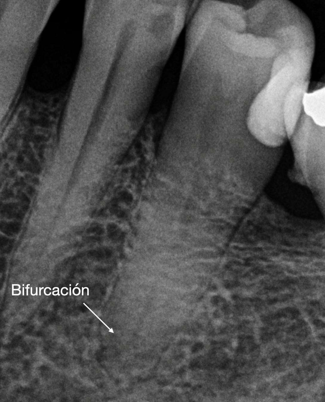 7 imgs
Bifurcación apical trabajada con limas manuales de pequeño calibre. El estudio de la radiografía inicial es clave para planificar el abordaje y no crear escalones con las limas rotatorias.
11, 12, 13 y 14 de noviembre de 2026 · Sevilla, España · Presencial
Endodoncia y Cirugía Apical · Endodoncia Avanzada
The Journal of the American Dental Association
Q3 · IF 1.332Autores · Nuria Castells-Gasa, Óliver Valencia de Pablo, Óscar Alonso-Ezpeleta, Silvia Herrero-Hernández, Alejandro Peña López, Antonio Conde, Ruth Perez-Alfayate, Giampiero Rossi-Fedele, Roberto Estévez
BACKGROUND: The palatogingival groove is a developmental anomaly that originates in the central fossa and extends along the root surface, often resulting in periodontal, endodontic, or combined endodontic and periodontal involvement. The authors present a case of a type III palatogingival groove associated with a periodontal defect and a sinus tract managed successfully through surgical and regenerative treatment. CASE DESCRIPTION: A woman sought treatment at Clínica Dental ZOE in Zaragoza, Spain, with the chief symptoms of pain and swelling associated with tooth no. 7. Clinical and radiographic findings supported the diagnosis of type III palatogingival groove in the presence of a normal pulp. The additional root was resected, the periradicular lesion was curetted, and guided bone regeneration procedures were carried out. Long-term recall visits through 3 years confirmed successful retention of the tooth and periodontal regeneration in the absence of pathosis. PRACTICAL IMPLICATIONS: The authors describe an unusual clinical manifestation and emphasize the role of accurate diagnosis leading to avoidance of unnecessary endodontic treatment, providing a reference for similar clinical situations. Considering the broad spectrum of manifestations of palatogingival grooves, a case-by-case multidisciplinary approach to management is recommended.
Australian Endodontic Journal
Q3 · IF 1.202Autores · Yoseif Haddad, Wing Chuan Chan, William Nguyen Ha, Antonio Conde, Roberto Estévez, Alejandro Peña López, Georgia Charlotte Gates, Giampiero Rossi‐Fedele
This study assessed the antimicrobial effect of sodium hypochlorite (NaOCl) mixtures combined with Keratobacter (KB) using an engineered biofilm root canal model. Clinical and reagent grade NaOCl were mixed with KB (9:1-vol/vol) to assess pH values over 1 min to select the ideal solution with a pH just below the pKa of hypochlorous acid. The samples were randomly divided into five groups: 1% and 4% NaOCl reagents, a mixture of NaOCl:KB using 1% and 4% NaOCl reagents and distilled water. Outcome measures were colony-forming units (CFUs/mL) and positive/negative cultures. No significant differences were observed in the pairwise comparisons between 1%, 4% NaOCl and 4% NaOCl+KB for the outcome CFUs/mL. Only 4% NaOCl presented with negative cultures in all samples, whereas 1% NaOCl and 4% NaOCl+KB had similar results (54% vs. 40%). The addition of KB has a limited effect on the antimicrobial efficacy of 4% NaOCl in this laboratory model.
Journal of Endodontics
★ Alto impacto · Q1 · IF 3.424Autores · Alejandro Peña López, Antonio Conde, Roberto Estévez, Óliver Valencia de Pablo, Giampiero Rossi‐Fedele, Rafael Cisneros
The aim of this study was to evaluate porcine palatal mucosa dissolution by sodium hypochlorite (NaOCl) with or without an auxiliary dissolving agent containing glycocholic acid and a mixture of surfactants (Keratobacter [KB]; Saint Joseph DID, Valencia, Spain). One hundred forty samples were obtained from porcine palatal mucosa and weighed using a high-precision balance. The samples were randomly divided into 4 experimental groups (n = 35) based on the test solution used: distilled water (the negative control), CanalPro NaOCl 6% (Coltene Whaledent, Altstätten, Switzerland), KB, and a 9:1 vol/vol mixture of NaOCl with KB (NaOCl + KB). After 5, 10, 15, and 20 minutes of immersion in the solutions at 27°C ± 1°C, the samples were weighted by a blinded assessor. The intergroup weight at the different time points was statistically analyzed using the analysis of variance test with the Bonferroni posttest. All test groups presented with tissue dissolution although complete dissolution did not occur in any sample. The largest percent in weight reduction occurred between time points (t) = 0 minutes and t = 5 minutes for the NaOCl + KB group (22.5%) followed by KB (18.5%) for the same time period. NaOCl presented with similar tissue dissolution activity during the different time points, ranging from 7.8% (t = 10 minutes-t = 15 minutes) to 6.8% (t = 15 minutes-t = 20 minutes). Significant weight differences were found among the different experimental groups after 5, 10, and 15 minutes of incubation, with the only exception being KB versus NaOCl + KB. No significant differences were found when comparing the test groups at t = 20 minutes. The addition of KB to NaOCl increased porcine palatal mucosa dissolution in vitro.
Endodoncia y Cirugía Apical
Alicante · España
Endodoncia y Cirugía Apical
Santa Cruz de Tenerife · España
Endodoncia y Cirugía Apical
Madrid · España
Endodoncia y Cirugía Apical
Madrid · España
Endodoncia y Cirugía Apical
Zaragoza · España
Endodoncia y Cirugía Apical
Huesca · España