Cirugía Oral e Implantología
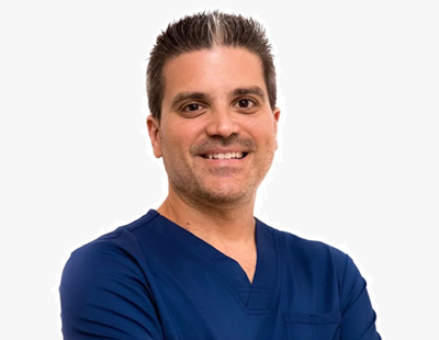
Toni Flichy centra su práctica en Cirugía Oral e Implantología. Ejerce en Valencia. Su formación de base procede de Universitat de Barcelona en 2006.
A lo largo de su trayectoria, ha llegado al nivel de doctorado. Su título de posgrado más reciente es el Doctorado en Odontología (Universidad de Valencia, 2014).
La docencia ocupa la otra mitad de su tiempo: imparte clase en Universidad de Valencia, SCOE y Universidad Rey Juan Carlos, en programas como Máster de Formación Permanente en Cirugía Oral e Implantología y Postgrado de Implantología Oral.
En Doctor&Cols dirige e imparte 9 formaciones — entre ellas «Diseño interdisciplinar de la sonrisa: integración de la odontología restauradora y la cirugía estética periodontal» y «Curso intensivo en manejo de tejidos blandos y mucogingival» — en Clínica Dental Teresa Alegre · Plaza del Poble 8, bajo, Silla (Valencia) y Barcelona · Calle Còrcega 474 y firma 21 publicaciones científicas en revistas como Journal of Clinical Medicine e International Journal of Immunopathology and Pharmacology.
Explora su trayectoria
Doctorado en Odontología
Doctorado · Universidad de Valencia · Valencia, España
Máster de Formación Permanente en Cirugía Oral e Implantología
Máster · Universidad de Valencia · Valencia, España
Grado / Licenciado en Odontología
Grado · Universitat de Barcelona · Barcelona, España
Sin experiencia registrada
Profesor colaborador · Máster de Formación Permanente en Cirugía Oral e Implantología
Universidad de Valencia · Valencia, España
Profesor colaborador · Postgrado de Implantología Oral
SCOE · Barcelona, España
Profesor colaborador · Máster de Formación Permanente en Cirugía Bucal e Implantología
Universidad Rey Juan Carlos · Madrid, España
Profesor asociado · Máster periodoncia quirúrgica y periimplantaria
Universidad Católica San Antonio de Murcia (UCAM) · Málaga, España
Módulo 1: 27-29 de enero de 2027 · Módulo 2: 24-26 de febrero de 2027 · Barcelona · Presencial
Cirugía oral · Regeneración ósea
21, 22 y 23 de abril de 2027 · Barcelona · Calle Còrcega 474 · Presencial
Cirugía y Periodoncia · Cirugía Mucogingival
16, 17 y 18 de junio de 2027 · Barcelona · calle Bruc 29 y ESIRO Barcelona · Presencial
Cirugía y Periodoncia · Cirugía Mucogingival
30 de septiembre y 1 y 2 de octubre de 2026 · Barcelona · ESIRO Barcelona, calle Còrcega 474 · Presencial
Cirugía y Periodoncia · Periimplantitis
24 y 25 de septiembre de 2026 (jueves de 13:00 a 21:00 y viernes de 9:30 a 21:00) · Silla (Valencia) · Clínica Dental Teresa Alegre, Plaza del Poble 8, bajo · Presencial
15 y 16 de octubre de 2026 · Barcelona · ESIRO Barcelona, calle Córcega 474 · Presencial
Cirugía y Periodoncia · Cirugía Avanzada y Regenerativa
Del 26 al 30 de octubre de 2026 · São Paulo, Brasil · Universidad FACOP · Presencial
Cirugía y Periodoncia · Cirugía Avanzada y Regenerativa
27 y 28 de noviembre de 2026 · Clínica Dental Teresa Alegre · Plaza del Poble 8, bajo, Silla (Valencia) · Presencial
Armonización Orofacial · Diseño de Sonrisa
18, 19 y 20 de noviembre de 2026 · Barcelona · calle Bruc 29 y ESIRO Barcelona · Presencial
Cirugía y Periodoncia · Cirugía Guiada
Journal of Clinical Medicine
Autores · Antonio Juan Flichy‐Fernández, Miguel Padial‐Molina, Natividad Martín‐Morales, T. Alegre‐Domingo, Miguel Peñarrocha Diago, Francisco O’Valle, Pablo Galindo‐Moreno
Background: To compare the histological and histomorphometrical outcomes after sinus floor elevation using an anorganic bovine bone biomaterial or a biphasic calcium phosphate biomaterial. Material and Methods: Patients who needed maxillary sinus elevation were included in this study. A total of 68 implant sites were evaluated from a total of 42 patients. Twenty patients were treated with anorganic bovine bone, while 22 were treated with biphasic calcium phosphate biomaterial. Morphological and morphometrical studies were performed on the bone samples collected during implant placement. Results: Both biomaterials induced similar relative areas of mineralized tissue overall, particularly if only the area of grafted bone was considered. In turn, a higher proportion of non-mineralized tissue was observed in cases of biphasic calcium phosphate biomaterial with less area of remnant biomaterial particles. None of the implants failed at one year of follow-up. Conclusions: Although both biomaterials induce a similar amount of bone formation, the histopathological characteristics of the grafts are different, with a greater proportion of scar connective tissue with the biphasic calcium phosphate biomaterial.
International Journal of Immunopathology and Pharmacology
Autores · Carla Borrell García, Marta Ribelles Llop, María Ángeles García Esparza, Antonio Juan Flichy‐Fernández, Laura Marqués Martínez, Regina Izquierdo Fort
To assess the effects of a probiotic upon oral health indices in adolescents and to establish relationships between these indices and dietary habits and oral hygiene. Twenty-seven adolescents between 12 and 18 years of age were randomized into two groups. The study group received tablets containing Lactobacillus reuteri DSM 17938/ ATCC 5289 for 28 days, while the control group received tablets without any bacteria. Streptococcus mutans, Lactobacillus sp., and salivary pH were assessed at baseline and at 7, 14, 21, 28, and 45 days. The plaque, gingivitis, and bleeding indices were recorded at baseline and at 14, 28, and 45 days. Dietary and oral hygiene habits were also evaluated by means of a questionnaire. A less marked rise in S. mutans was recorded in the study group. Improvements were observed in terms of plaque, gingivitis, and bleeding, though statistical significance was not reached. Oral pH increased in the study group, though not to a significant degree. Poorer eating habits were significantly correlated to increased plaque. The study parameters decreased with the two strains of L. reuteri DSM 17938 and ATCC PTA 5289, though the results failed to reach statistical.
Materials
Autores · Eugenio Velasco‐Ortega, Antonio Juan Flichy‐Fernández, Miquel Punset, Álvaro Jiménez-Guerra, José María Manero, Javier Gil
Sixty-four fractured commercially pure titanium (cp-Ti) narrow dental implants (NDIs) with similar macrogeometry and connection designs were studied after different implantation times in humans in order to determine their reliability and to evaluate the causes of the fracture. These NDIs were compared with other similar implants, made with alloyed titanium with 15% Zr and with 12% strained titanium. Original implants were tested under static and fatigue conditions, simulating the tri-axial loads in the mouth by means of a Bionix hydraulic test machine. Fractography was studied using field-emission scanning electron microscopy (FSEM). The results showed that cp-Ti NDI exhibits low strength for mechanical cycling, and the alloyed Ti and strained titanium increase the mechanical strength, guaranteeing long term mechanical behavior. NDIs fractured due to fatigue, and, in some cases, the presence of cracks in the original NDIs quickly led to fracture. These cracks were attributed to plastic deformation during machining were found to be exacerbated due to acid etching in the passivation process. All cases of fracture were cp-Ti dental implants due to the low fatigue limit. The results show that, when titanium is alloyed or cold-worked, the fatigue limit is higher than cp-Ti. This in vitro research will help clinicians to select a better NDI system for safer treatment.
Clinical Implant Dentistry and Related Research
Autores · Antonio Juan Flichy‐Fernández, Juan Antonio Blaya-Tárraga, Francisco O’Valle, Miguel Padial‐Molina, Miguel Peñarrocha Diago, Pablo Galindo‐Moreno
BACKGROUND: Poly (lactic-co-glycolic acid) (PLGA) is widely used for the development of delivery systems for drugs and therapeutic biomolecules in tissue engineering applications. Particles of biphasic calcium phosphate can be covered by PLGA to change their manipulating characteristics. PURPOSE: Aim of this study was to investigate the radiological and histomorphometric results of the use of PLGA-coated biphasic calcium phosphate granules in sinus floor elevation and to analyze the underlying molecular processes by immunohistochemical staining. MATERIALS AND METHODS: A randomized clinical study was designed to include patients in need of sinus floor elevation. Patients were assigned to receive either PLGA-coated biphasic calcium phosphate particles (group I) or the equivalent but noncoated particles (group II). Cone beam computed tomography (CBCT) scans were performed before and 6 months after the procedure to assess the bone height gain. At the time of implant placement, bone core biopsies were obtained at the site of implant placement. Histological sections were subjected to histomorphometric and immunohistochemical evaluation of differentiation markers (Musashi-1 [MSI1]). RESULTS: No statistically significant differences were observed between groups for the radiologic parameters. No differences were observed histologically or histomorphometrically. However, PLGA-coated particles (group I) were more colonized by MSI1-positive osteoblast precursors (P = 0.0001, chi-squared test) and were penetrated by more CD34-positive vascular structures (P = 0.001, chi-squared test) than noncoated particles (group II). CONCLUSIONS: PLGA-coated particles are associated with more MSI11-positive cells and more extensive microvascularization than noncoated particles.
Journal of Periodontal Research
★ Alto impacto · Q1 · IF 6.385Autores · A. J. Flichy-Fernández, J. Ata-Ali, T. Alegre-Domingo, E. Candel-Martí, F. Ata-Ali, J. R. Palacio, M. Peñarrocha-Diago
Los probióticos crean un biofilm que protege los tejidos orales frente a la acción de bacterias periodontopatógenas. El objetivo del estudio fue evaluar los efectos del probiótico oral Lactobacillus reuteri Prodentis sobre la salud periimplantaria de pacientes edéntulos con implantes dentales y mucositis periimplantaria, comparándolos con implantes sin enfermedad periimplantaria. Ensayo clínico aleatorizado, doble ciego, controlado.
BMC Oral Health
★ Alto impacto · Q2 · IF 3.535Autores · Javier Ata-Ali, Antonio Juan Flichy-Fernández, Teresa Alegre-Domingo, Fadi Ata-Ali, Jose Palacio, Miguel Peñarrocha-Diago
Debido al aumento mundial de los tratamientos con implantes, la incidencia de enfermedad periimplantaria está en crecimiento. El fracaso tardío del implante es consecuencia de la incapacidad para mantener la osteointegración, cuya causa más importante es la periimplantitis. El objetivo del estudio fue analizar los aspectos clínicos, microbiológicos e inmunológicos en el fluido crevicular periimplantario (PISF) de pacientes con implantes sanos y con periimplantitis.
Journal of Investigative and Clinical Dentistry
Q2 · IF 0Autores · Javier Ata-Ali, Antonio Juan Flichy-Fernández, Teresa Alegre-Domingo, Fadi Ata-Ali, Miguel Peñarrocha-Diago
El objetivo del estudio fue investigar cómo el tabaquismo severo influye en los parámetros clínicos, microbiológicos y de respuesta del huésped en el fluido crevicular periimplantario de pacientes con implantes dentales sanos. Se evaluaron 29 individuos con 74 implantes: 20 implantes en fumadores severos y 54 en no fumadores. Se analizaron índice gingival modificado, índice de placa modificado y profundidad de sondaje, además de bacterias periodontopatógenas (Tannerella forsythia, Treponema denticola y otras).
The International Journal of Oral & Maxillofacial Implants
Autores · Javier Ata‐Ali, Antonio Juan Flichy‐Fernández, Fadi Ata‐Ali, María Peñarrocha‐Diago, Miguel Peñarrocha Diago
PURPOSE: To analyze the clinical, microbiologic, and host response characteristics (interleukins 1β and 6) in the peri-implant sulcus fluid (PISF) of patients with healthy dental implants and with peri-implant mucositis. MATERIALS AND METHODS: Clinical parameters (modified Gingival Index [mGI], modified Plaque Index [mPI], probing pocket depth [PPD], and absence or presence of radiologic bone loss) were recorded, and PISF samples were obtained from peri-implant sites showing mucositis as well as healthy sites. The periodontopathogenic bacteria Tannerella forsythia (Tf), Treponema denticola (Td), and Porphyromonas gingivalis (Pg) were evaluated, together with the total bacterial load (TBL) and the interleukin (IL) 1β and IL-6 values. RESULTS: The study population consisted of 34 individuals, and 77 dental implants were evaluated during the study (23 mucositis and 54 healthy peri-implant sites). The mGI, mPI, and PPD scores of the peri-implant mucositis group were significantly greater than in the healthy group. No differences in detection frequency were found for putative periodontal pathogens and TBL between the healthy peri-implant sites and mucositis sites. The mucositis group showed a significantly greater expression of IL-6 than the healthy group (P < .05). Although IL-1β was increased in the mucositis group, there was no statistically significant difference versus the healthy implant group. CONCLUSIONS: An analysis was made of the clinical, microbiologic, and host response characteristics in implants with peri-implant mucositis, establishing comparisons with healthy implants. In the patients studied, bacterial plaque induced an inflammatory response that can lead to the development of peri-implant mucositis. Adequate plaque control is therefore able to increase peri-implant health, avoiding the risk of future complications. No specific association to the studied bacterial species was established.
Medicina Oral, Patología Oral y Cirugía Bucal
Q2 · IF 2.165Autores · Hilario Pellicer-Chover, David Peñarrocha-Oltra, Leticia Bagán, Antonio J. Flichy-Fernández, Luigi Canullo, Miguel Peñarrocha-Diago
Ensayo clínico prospectivo, aleatorizado, simple ciego, que comparó salud periimplantaria, pérdida ósea marginal y éxito de implantes inmediatos vs diferidos en rehabilitaciones full-arch fijas. 15 pacientes, 144 implantes (76 diferidos, 68 inmediatos). A los 12 meses no hubo diferencias estadísticamente significativas en éxito ni pérdida ósea marginal (0,54 ± 0,39 mm inmediatos vs 0,66 ± 0,25 mm diferidos; p=0,201). Ningún implante fracasó.
Clinical Oral Implants Research
★ Alto impacto · Q1 · IF 3.681Autores · M. A. Peñarrocha-Diago, A. J. Flichy-Fernández, R. Alonso-González, D. Peñarrocha-Oltra, J. Balaguer-Martínez, M. Peñarrocha-Diago
Objective: To carry out a comparative study of two implants with different neck features and prostheses platform connection (machined with external connection and rough-surfaced with switching platform) upon peri-implant marginal bone loss, before and after functional loading. Material and methods: A randomized, prospective radiological study was made. Eighteen totally edentulous patients were selected. Subjects were divided into two groups according to the type of implant neck used: (a) Osseous®, with machined surface, without microthreads, external connection, and without platform switching; and (b) Inhex®, with treated surface, microthreads, internal connection, and platform switching. Mesial and distal marginal bone loss was measured. Implant success was assessed according to the criteria of Buser. Control timepoints were as follows: (a) at implant placement; (b) at prosthesis placement; (c) 6 months after loading; (d) 12 months after loading. Results: Fifteen patients that received 120 dental implants were included: 47% Osseous® group and 53% Inhex® group. Global mean marginal bone loss with Osseous® was 0.27 ± 0.43 mm and 0.38 ± 0.51 mm as determined 6 and 12 months after prosthetic loading, respectively, whereas in the case of Inhex® was 0.07 ± 0.13 and 0.12 ± 0.17 mm. These differences were statistically significant (P = 0.047). Difference between Osseous® and Inhex® in maxilla (P = 0.272) and mandibular (P = 0.462) bone loss were not statistically significant. Conclusions: Bone loss after 6 and 12 months proved statistically significant between two groups, with comparatively greater loss in the case of Osseous® implants vs. Inhex® implants. Regardless the heterogeneity of the two groups (neck shape, microthreads, surface texture), the implant–abutment connection appears to be a significant factor on peri-implant crestal bone levels. Anyway, in both groups, the values obtained were within normal ranges described in the literature.
Journal of Clinical and Experimental Dentistry
Q3 · IF 1.368Autores · A. J. Flichy-Fernández, S. González-Lemonnier, J. Balaguer-Martínez, D. Peñarrocha-Oltra, M. A. Peñarrocha-Diago, J. V. Bagán-Sebastián
Osteonecrosis of the jaw (ONJ) may appear following certain oral surgery procedures in patients treated with oral bisphosphonates (OB). Guidelines for the treatment of these patients were set out in the American Association of Oral and Maxillofacial Surgeons (AAOMS) Position Paper on Bisphosphonate-Related Osteonecrosis of The Jaws (Position Paper) and Approved by the Board of Trustees in September 2006. For the AAOMS the placement of implants in these patients is not contraindicated. In addition, the serum C-terminal telopeptide bone suppressor marker (CTX) test is available to determine the risk of ONJ. A case is presented of ONJ in a patient with 6 months of OB discontinuation (“drug holiday”) before dental implant placement (following the guidelines of the AAOMS) and with no risk of osteonecrosis according to the serum CTX value (340 pg/ml). The wound healed favorably with complete healing at 7 months. In this case, the serum CTX test must be questioned as to its predictive value of ONJ, and more reliable markers of this risk are needed. Key words: Bisphosphonates, dental implants, bone necrosis, serum CTX.
Medicina Oral, Patología Oral y Cirugía Bucal
Q2 · IF 2.165Autores · Antonio J Flichy-Fernández, Teresa Alegre-Domingo, Sandra González-Lemonnier, José Balaguer-Martínez, María Peñarrocha-Diago, Yolanda Jiménez-Soriano, David Peñarrocha-Diago, José V Bagán-Sebastián
Objectives: To determine whether there is a relationship between the total BP dose administered and the variations in serum CTX concentration. Study design: The study included 50 patients requiring dental implant surgery and treated with oral BPs, seen in an Oral Surgery and Implantology Unit between January 2007 and June 2009. The patients were divided into two groups: those in which the medication was not suspended before obtaining the laboratory test sample, and those patients referred from other dental clinics in which BPs was suspended before reporting to our Unit. The total drug dosage administered and the total dose per kilogram body weight were evaluated for comparison with serum CTX. The data obtained were correlated to the osteonecrosis risk table developed by Marx et al. in 2007. Results: There were no significant differences between the two groups in relation to the total administered dose and the dose in mg/kg b.w. Likewise, in both groups no relationship was observed between the serum CTX value and the total administered dose or the dose in mg/kg b.w. No differences were found between the two patient groups regarding chemical osteonecrosis risk based on the criteria of Marx et al. Conclusions: No relationship was observed between the oral BP dose administered (total dose or expressed in mg/kg b.w.) and serum CTX concentration, and suspension of the medication did not influence the serum CTX levels.
Journal of Clinical and Experimental Dentistry
Q3 · IF 1.368Autores · Fadi Ata-Ali, Javier Ata-Ali, Antonio J Flichy-Fernández, José V Bagan
The literature describes an increasing presence of bisphosphonate-induced osteonecrosis of the jaws (ONJ), characterized by the exposure for over 8 weeks of necrotic bone in the maxillofacial region, after bisphosphonate therapy, in the absence of prior maxillary radiotherapy. The present literature review examines the etiopathogenesis, risk factors, clinical forms, diagnosis, treatment and prevention of bisphosphonate-induced ONJ. In addition, a review is made of all the series involving over 15 patients diagnosed with this disorder between 1 January 2011 and 15 May 2011. A PubMed-Medline search was carried out with the following key words: “bisphosphonates” and “osteonecrosis”. The appearance of osteonecrosis is a serious complication, with an increasing incidence, that affects patient quality of life and causes important morbidity. All patients treated with bisphosphonates are at risk of developing osteonecrosis as a result of such medication. This potential complication therefore should be explained to the patient by both the prescribing physician and the dental surgeon in charge of oral treatment, with the obtainment of informed consent in all cases.
Medicina Oral, Patología Oral y Cirugía Bucal
Q2 · IF 2.131Autores · Ata-Ali J, Candel-Marti ME, Flichy-Fernández AJ, Peñarrocha-Oltra D, Balaguer-Martinez JF, Peñarrocha Diago MA
Introduction: Peri-implantitis is a late complication of dental implant treatment, induced by microbiological changes. Since the disorder is frequent, a review is indicated of the microorganisms that influence it and of the existing treatment options. Objective: To conduct a literature review of the microbiota associated to peri-implantitis and the existing treatment options. Material and Method: A PubMed literature search was made of the studies on the microbiota associated to dental implants in healthy patients and patients with peri-implantitis, as well as of the latest treatment developments, using the following key words: "peri-implantitis AND microbiota", "periimplantitis AND microbiota", "peri-implantitis AND treatment", and "periimplantitis AND treatment". Only clinical studies in humans were considered. The following criteria were applied for including articles in the analysis: a) for the peri-implant microbiota, the search limits were human studies after the year 2000; and b) for the treatment of peri-implantitis, the search limits were randomized and controlled clinical trials (RCTs) in humans, with a minimum follow-up of 4 months, and publication after the year 2000. Results: A total of 18 articles were selected in relation to peri-implant microbiota, and 13 in relation to the treatment of peri-implantitis (8 involving nonsurgical mechanical treatments and 5 surgical procedures). Conclusions: Evaluation of the literature has shown the microbiota associated to peri-implantitis to be more complex than that found under healthy peri-implant conditions – the main flora consisting of anaerobic gramnegative bacteria. No clear criteria have been identified for the diagnosis and treatment of peri-implantitis.
Medicina Oral, Patología Oral y Cirugía Bucal
Q2 · IF 2.131Autores · Ata-Ali J, Flichy-Fernández AJ, Alegre-Domingo T, Candel-Marti ME, Peñarrocha D, Balaguer-Martinez JF, Peñarrocha MA
Objective: To evaluate the presence within the peri-implant sulcus of Tannerela forsythia (Tf), Porphyromonas gingivales (Pg), Treponema denticola (Td) and Aggregatibacter actinomycetemcomitans (Aa), and relate these bacteria to the peri-implant crevicular fluid volume (PICFV). Material and Method: A prospective and cross-sectional clinical case series study was made. For the measurement of crevicular fluid, use was made of the Periotron® 8000 (Proflow Incorporated. New York, USA), measuring the volume in Periotron units (PU). For the detection of periodontopathogenic bacteria we used the IAI-PadoTest 4.5 (IAI Inc., IAI Institute, Zuchwil, Switzerland) – a system for the detection of Tf, Pg, Td and Aa based on the use of RNA arrays. Results: We included 34 patients (19 females and 15 males) with a mean age of 56.4 years. Of these subjects, 30.8% were smokers and 69.2% non-smokers. Out of a total series of 213 implants, we analyzed the crevicular fluid and microbiota in 90 implants. A total of 16.5% of the implants presented mucositis, while 83.5% were in healthy peri-implant conditions. The microbiological study revealed the presence of Tf in 17.1% of the implants, Pg in 9.3%, Td in 13.6%, in Aa in none of the implants. The mean Periotron reading was 93.4 PU (range 12-198 PU). A statistically significant (p<0.05) relationship was observed between PICFV and the total percentage bacteria (Tf, Pg and Td) – with a strong association between the Td levels and smoking (p<0.01). In the implants with mucositis, the concentration of Pg and Td was greater. Conclusions: In the implants studied, the subgingival peri-implant microbiota was characterized by low levels of Pg, Tf, Td, and none of the patients proved positive for Aa. These bacteria showed a positive correlation to crevicular fluid volume, and a statistically significant relationship was observed between Td and smoking.
Medicina Oral, Patología Oral y Cirugía Bucal
Q2 · IF 2.131Autores · Candel-Martí ME, Flichy-Fernández AJ, Alegre-Domingo T, Ata-Ali J, Peñarrocha-Diago MA
Introduction: A study is made of the usefulness of cytokines (such as interleukin-6 (IL-6), interleukin-8 (IL-8), interleukin-10 (IL-10) and interleukin-12 (IL-12)) as markers of periimplant disease (mucositis and periimplantitis). An increase in the levels of these cytokines in dental implant crevicular fluid may give rise to a lack of osteointegration, bone loss or implant failure. Objective: To review the literature relating IL-6, IL-8, IL-10 and IL-12 levels to dental implant surgery and periimplantitis. Material and Method: A PubMed literature search was made of articles in English and Spanish, using the key words "cytokine and dental implants", "cytokine and periimplantitis", "IL-6, IL-8, IL-10, IL-12 and dental implants", "IL-6, IL-8, IL-10, IL-12 and periimplantitis". Fourteen articles were found and classified into two groups relating interleukin levels to: a) periimplant disease; and b) their influence upon dental implant osteointegration without periimplant disease. Conclusions: An increase in interleukin levels is observed in patients with periimplant disease, though there is controversy over the effect of interleukins in crevicular fluid and periimplantitis in relation to implant failure or the development of periimplant disease.
Journal of Clinical and Experimental Dentistry
Autores · L. Gonzalez-Munoz, AJ. Flichy-Fernandez, Javier Ata‐Ali, A. Pascual-Moscardo, MA. Penarrocha-Diago
Objective: To review the literature on ozone therapy in oral health, as assessed by different clinical and bacteriological parameters. Material and methods: A PubMed literature search was made using the key words "ozone dental", and establishing as limits "randomized controlled trial" and "dental journal". Thirteen articles were identified, with access to only 6 of them. Results: Four studies used ozone for the treatment of caries. One study examined its effect upon dental hypersensitivity, while another evaluated the efficacy of ozone as a tooth whitening technique. Five studies explored the bacteriological actions of ozone therapy in reference to different types of bacteria.
Journal of Clinical and Experimental Dentistry
Autores · AJ. Flichy-Fernandez, Javier Ata‐Ali, C. Palma-Carrio, D Penarrocha-Oltra, J. Alejandro Conejero, M. Penarrocha
Introduction: Following dental implant loading, marginal bone loss after one year must be evaluated to check correct maintenance of the bone levels. Objectives: To assess implant treatment success and quantify marginal bone loss 6 and 12 months after loading. Material and method: Sixty-one MIS implants with a 1.8 mm machined neck were placed in 26 patients. Implant success was based on the criteria of Buser. Radiological controls were made 6 and 12 months after loading, measuring bone loss mesial and distal. Results: Twenty-two patients with 56 implants were included: 32 in the maxilla and 24 in the mandible. Two implants failed in two patients during the osseointegration phase (both in the maxilla), yielding an implant success rate of 96.4%. After 6 months, bone loss was 0.801.04 mm mesial and 0.731.08 mm distal, while after 12 months bone loss was 0.921.02 mesial and 0.871.01 distal. Conclusions: Bone loss 6 and 12 months after machined neck implant placement was within the normal ranges described in the literature.
Medicina Oral, Patología Oral y Cirugía Bucal
Q2 · IF 2.131Autores · Flichy-Fernández AJ, Alegre-Domingo T, Peñarrocha-Oltra D, Peñarrocha-Diago M
Probiotics have been found to be beneficial to host health. Their primary use in medicine has been for the management of intestinal tract problems. In recent years, probiotics have been used as a treatment to promote oral health. The aim of the present study was to review published studies regarding probiotics and their effects on the oral cavity. Studies reporting the anticariogenic effects of probiotics, their use in the treatment of periodontal disease, a reduced crevicular fluid volume and cytokine content, as well as their use in the treatment of halitosis and Candida albicans were identified. Studies assessing residence time of probiotics in the oral cavity were also selected for retrieval. Most authors concluded that the use of oral probiotics was associated with an improvement in oral health, including a significantly reduced level of cariogenic and periodontal pathogens and a lower crevicular fluid volume and cytokine concentration.
Medicina Oral, Patología Oral y Cirugía Bucal
Q2 · IF 2.131Autores · Flichy-Fernández AJ, Balaguer-Martínez J, Peñarrocha-Diago M, Bagán JV
Osteonecrosis of the jaw has been described in patients taking bisphosphonates after oral surgery procedures, including the placement of dental implants. This review is an update of the relationship between bisphosphonates and dental implants. Results obtained by different authors are compared, contrasting earlier studies where an improvement in implant osseointegration using bisphosphonates was observed, with ones where statistically significant differences were found, and more recent studies disagreeing with the use of bisphosphonates for causing necrosis of the jaw. The differing results obtained between animal studies and the situation observed in humans may be due to a short medication and follow-up period, as well as to the existence of few research studies where dental implants are placed in the oral cavity. Currently, dental implants are contraindicated in patients being treated with intravenous bisphosphonates. In 2007, the American Association of Oral and Maxillofacial Surgeons suggested guidelines for patients treated with oral bisphosphonates, based on the clinical situation of the patient and the length of treatment with the drug, and that greater caution prior and subsequent to surgery should be taken for three years after treatment. All patients treated with bisphosphonates must have the risk of possible loss of implants and the risk of suffering a bony necrosis of the operated jaw explained to them, and give their informed consent prior to dental implant surgery.
Medicina Oral, Patología Oral y Cirugía Bucal
Q2 · IF 2.131Autores · Martínez-Ricarte J, Faus-Matoses V, Faus-Llácer VJ, Flichy-Fernández AJ, Mateos-Moreno B
Dentinal sensitivity is a clinical condition of some importance, particularly in periodontal patients. Symptoms appear on applying a triggering stimulus to the exposed dentine - the particularity being that the pain is similar to that of other dental disorders of different etiology and treatment. Hence the importance of a correct differential diagnosis. The main problem not only in clinical practice when treating the disorder, but also in designing studies for the objective evaluation of dentinal sensitivity, is the difficulty of standardizing, evaluating and interpreting the clinical condition in its different degrees. Thus, consensus in designing and evaluating studies of dentinal sensitivity would facilitate our understanding of its etiology, and the assessment of possible treatments. Such studies may center their methodology on individual patient response or on the nature of the triggering stimulus. The present study provides an updated and global view of the disorder, and reviews the basic protocol for the objective assessment of dentinal sensitivity.
Cirugía Oral e Implantología
Sevilla · España
Cirugía Oral e Implantología
Vizcaya · España
Cirugía Oral e Implantología
Almería · España
Cirugía Oral e Implantología
Madrid · España
Cirugía Oral e Implantología
Valencia · España
Cirugía Oral e Implantología
Sevilla · España