Cirugía Oral e Implantología

Markel Diéguez Pereira centra su práctica en Cirugía Oral e Implantología. Ejerce en Vizcaya. Cursó sus estudios de Odontología en Universidad del País Vasco en 2010.
Con el tiempo, ha sumado formación específica en periodoncia y ha llegado al nivel de doctorado. Su título de posgrado más reciente es el Doctorado en Odontología (Universidad de Oviedo, 2021). Ha pasado por Universidad de Oviedo y Universidad CEU Cardenal Herrera.
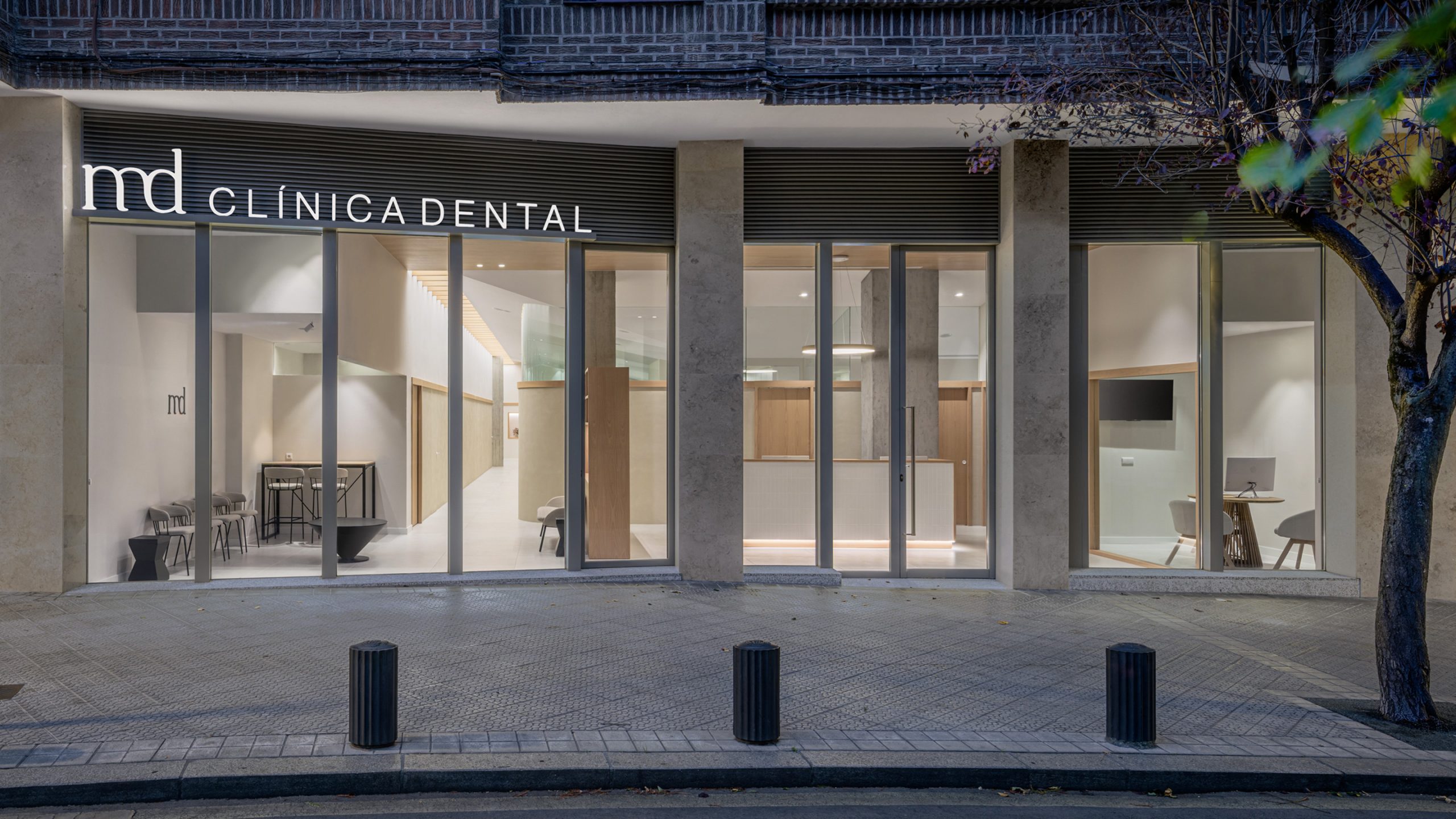
Su práctica diaria se desarrolla en Clínica dental MD, como cirujano/implantólogo (Vizcaya, España).
Además de la clínica, mantiene actividad de docencia en Universidad Europea Miguel de Cervantes (UEMC) y Universidad de Zaragoza, en programas como Experto Universitario en Implantología Oral y Máster Propio en Periodoncia e Implantología Oral.
Su aportación al colectivo: firma 24 publicaciones científicas en revistas como Journal of Functional Biomaterials y Journal of Clinical Medicine.
Explora su trayectoria
Doctorado en Odontología
Doctorado · Universidad de Oviedo · Oviedo, España
Máster de Formación Permanente en Cirugía Bucal, Implantología Bucofacial y Periodoncia
Máster · Universidad CEU Cardenal Herrera · Valencia, España
Especialización Universitaria Implantología Oral y Microcirugía
Especialista · Universidad del País Vasco · Leioa, España
Curso de Especialización en Implantología Oral
Curso · Universidad de Santiago de Compostela · A Coruña, España
Grado / Licenciado en Odontología
Grado · Universidad del País Vasco · Leioa, España
Clínica dental MD · Vizcaya, España
Profesor · Experto Universitario en Implantología Oral
Universidad Europea Miguel de Cervantes (UEMC) · Valladolid, España
Profesor colaborador · Máster Propio en Periodoncia e Implantología Oral
Universidad de Zaragoza · Zaragoza, España
Journal of Functional Biomaterials
★ Alto impacto · Q1 · IF 5.16Autores · Esteban Pérez-Pevida, Iván Monteagudo-Villalobos, David Chávarri-Prado, Alejandro Estrada-Martínez, Miguel Beltrán-Guijarro, Markel Diéguez-Pereira, Aritza Brizuela-Velasco
Coautores Doctor&Cols · Esteban Pérez Pevida, David Chávarri Prado, Alejandro Estrada Martínez, Miguel Beltrán Guijarro, Iván Monteagudo
Resonance frequency analysis (RFA) has been used as a diagnostic method to measure implant stability at all stages of healing. In addition to evaluating the status of the peri-implant marginal bone, it can also indicate the most appropriate time to load the implant. This in vitro study aimed to evaluate the efficacy of RFA as a diagnostic method for the detection of peri-implant marginal bone loss (MBL). Forty bone-level Klockner Vega implants were placed in a polyurethane block with elastic properties similar to those of the maxillary bone. The insertion torque and primary implant stability at the time of placement were measured using an RFA device. A circumferential peri-implant defect was created by removing the cortical bone portion in each implant using a trephine. The stability values were measured again using RFA. The stability values measured using RFA were lower after the creation of the circumferential peri-implant defect, indicating a statistically significant decrease in implant stability. The results of the study tend to show a relationship between peri-implant marginal bone loss and modifications in implant stability measured by RFA.
Journal of Clinical Medicine
★ Alto impacto · Q1 · IF 3.408Autores · Oier Montalbán-Vadillo, Esteban Pérez-Pevida, Iratxe Viteri-Agustín, David Chávarri-Prado, Alejandro Estrada-Martínez, Markel Diéguez-Pereira, Fernando Sánchez-Lasheras, Aritza Brizuela-Velasco
Coautores Doctor&Cols · Esteban Pérez Pevida, Alejandro Estrada Martínez, David Chávarri Prado
Background: Although xenografts have shown successful results in GBR procedures due to their osteoconductive properties, many authors have opted to add co-adjuvant drugs to favor osteogenesis and differentiate cells into an osteoblastic lineage. Metformin has been shown to have bone-protective properties, regulating osteoclast differentiation, as well as the ability to promote osteoblast mineralization and differentiation. The present study aimed to evaluate the effect of the local application of a 1% metformin solution on bone neoformation in the treatment of an experimental bone defect in a guided bone regeneration animal model with a particulated bovine hydroxyapatite xenograft with hyaluronate. Methods: With this purpose in mind, two critical defects with 8 mm diameter and 0.5 mm depth were created in eight male New Zealand rabbit calvarias. Titanium cylinders were fixed in each defect and filled with particulate hydroxyapatite of bovine origin and sodium hyaluronate, with sterile injectable saline added to the control group and sterile 1% metformin solution added to the test group. At 6 weeks, the animals were euthanized, and samples were obtained and prepared for histomorphometric analysis. Results: A higher percentage of new bone formation was observed in the metformin samples than in the control samples, both in the region closest to the animal's calvaria and in the most distal region analyzed. A higher average bone–biomaterial contact percentage was observed in the samples, with metformin in both the proximal and distal regions. There was no statistically significant difference in the mean value in either region in both parameters. Conclusion: The local application of a 1% metformin solution in an animal model of guided bone regeneration with particulate bovine hydroxyapatite and hyaluronate resulted in greater bone neoformation and xenograft osseointegration than in the control group.
Journal of Functional Biomaterials
★ Alto impacto · Q1 · IF 5.284Autores · Miguel Beltrán-Guijarro, Esteban Pérez-Pevida, David Chávarri‐Prado, Alejandro Estrada-Martínez, Markel Diéguez-Pereira, Fernando Sánchez Lasheras, Aritza Brizuela‐Velasco
Coautores Doctor&Cols · Miguel Beltrán Guijarro, Alejandro Estrada Martínez, Esteban Pérez Pevida, David Chávarri Prado
This study aims to analyse, using a finite element analysis, the effects of Ti-base abutment height on the distribution and magnitude of transferred load and the resulting bone microstrain in the bone-implant system. A three-dimensional bone model of the mandibular premolar section was created with an implant placed in a juxta-osseous position. Three prosthetic models were designed: a 1 mm-high titanium-base (Ti-base) abutment with an 8 mm-high cemented monolithic zirconia crown was designed for model A, a 2 mm-high Ti-base abutment with a 7 mm-high crown for model B, and a 3 mm-high abutment with a 6 mm-high crown for model C. A static load of 150 N was applied to the central fossa at a six-degree angle with respect to the axial axis of the implant to evaluate the magnitude and distribution of load transfer and microstrain. The results showed a trend towards a direct linear association between the increase in the height of the Ti-base abutments and the increase in the transferred stress and the resulting microstrain to both the prosthetic elements and the bone/implant system. An increase in transferred stress and deformation of all elements of the system, within physiological ranges, was observed as the size of the Ti-base abutment increased.
The International Journal of Periodontics & Restorative Dentistry
Q3 · IF 1.103Autores · David Chávarri-Prado, Andoni Jones, Esteban Pérez-Pevida, Markel Diéguez-Pereira, Alejandro Estrada-Martínez, Rodrigo Cabezón-Palacios
Coautores Doctor&Cols · Andoni Jones, Esteban Pérez Pevida, Alejandro Estrada Martínez, David Chávarri Prado
Sinus floor augmentation is one of the most common approaches to obtain sufficient bone availability for placing implants in cases with severe bone atrophy in the posterior maxilla. Several bone substitutes are indicated for sinus augmentation, but they may achieve different clinical outcomes. This study aims to compare bovine bone mineral (BBM) with freeze-dried bone allograft (FDBA) in a two-stage lateral window sinus grafting approach. Twenty patients received a lateral window sinus elevation with either FDBA or BBM. Postoperative graft height was measured with CBCT. Implants were placed 6 months later, at which time biopsy samples were taken for histologic analysis and new CBCT scans were performed to measure graft height. The mean height reduction at 6 months was 20.27% ± 4.94% for FDBA samples and 5.36% ± 2.41% for BBM samples. The histologic analysis revealed a mean ratio of newly formed bone of 43.70% ± 5.29% for the FDBA group and 38.11% ± 4.03% for the BBM group. The FDBA group also showed a higher amount of residual biomaterial (17.25% ± 10.10%) and connective tissue (14.63% ± 4.38%) compared to the BBM group (15.53% ± 5.42% and 13.11% ± 4.42%, respectively). The differences between groups were statistically significant for the height reduction and newly formed bone (P ≤ .05) but not for the amounts of residual biomaterial and nonmineralized connective tissue (P ≥ .05). Six months after performing a lateral window sinus elevation, the percentage of newly formed bone was significantly higher when using FDBA than when using BBM, although the graft height reduction was also significantly higher for the FDBA group.
The International Journal of Oral & Maxillofacial Implants
Q2 · IF 1.088Autores · Chávarri-Prado D, Brizuela-Velasco A, Diéguez-Pereira M, Chento-Valiente Y, Estrada-Martínez A, Pérez-Pevida E, Álvarez-Arenal Á
Coautores Doctor&Cols · Esteban Pérez Pevida, David Chávarri Prado, Alejandro Estrada Martínez
Purpose: The aim of this study was to measure and compare the retention degrees of different attachment systems before and after 1 year of mastication and removal/insertion simulations. Materials and methods: A 4 × 10-mm implant embedded in self-curing acrylic resin was placed inside a rectangular metal frame installed in a plastic container with artificial saliva at 37°C. Three different attachment systems were connected to the implants: Dalbo Classic, Locator (with blue, pink, and clear inserts), and Klockner Rotula. A loading test at 100 N and 1.25 Hz frequency was repeated 10 times for each attachment (changing the matrix component or plastic insert) until a total of 300,000 cycles and 1,095 removal/insertion sequences. The retention force was assessed at baseline until the end of the test (1 year) after 3-month intervals of simulation. One-way analysis of variance (ANOVA) and post hoc Tukey test (P < .05) were performed for statistical analyses. Results: Klockner Rotula showed the highest retention values at the beginning (69.6 ± 9.2 N; 7.1 ± 0.9 kg) and the end of the test (51.4 ± 5.4 N; 5.2 ± 0.6 kg), while Dalbo Classic showed the lowest values both at the beginning (13.1 ± 1.7 N; 1.3 ± 0.2 kg) and the end of the test (6.7 ± 1.6 N; 0.7 ± 0.2 kg). Locator with clear insert exhibited the lowest retention loss ratio (20.4%), while Locator with blue insert showed the highest (50.8%). At the end of the test, all attachment systems showed a significant decrease in retention force in comparison with the initial retention (P < .001). Conclusion: Locator attachments with clear and pink inserts and the Klockner Rotula attachment system showed retention forces > 20 N (2 kg) after 1 year of mastication and prosthesis insertion/removal simulation. All attachment systems showed a progressive loss of retention, which was higher for Locator attachments with blue and pink inserts and Dalbo Classic. The Klockner Rotula attachment system showed the highest retention values both at the beginning and the end of the test.
International Journal of Implant Dentistry
★ Alto impacto · Q1 · IF 4.405Autores · Markel Diéguez-Pereira, David Chávarri‐Prado, Iratxe Viteri-Agustín, Oier Montalbán-Vadillo, Esteban Pérez-Pevida, Aritza Brizuela‐Velasco
Coautores Doctor&Cols · Esteban Pérez Pevida, David Chávarri Prado
BACKGROUND: Implantoplasty reduces both implant diameter and the thickness of its walls, subsequently reducing the ability of the implant to resist fracture in response to functional load. In combination with an increase in the crown-implant ratio due to bone loss, this could increase the lever effect, which in presence of high masticatory forces or parafunctional habits, could lead to complications such as fracture of the implant or loosening of the prosthetic screw. OBJECTIVES: To determine the elastic limits of internal connection, dental implants of different designs and diameters after an implantoplasty. MATERIALS AND METHODS: This in vitro study included 315 tapered internal connection titanium dental implants, the threads of which were removed with an industrial milling machine-for standardized implantoplasty (IMP1; n = 105)-or with the conventional approach-manually, using high-speed burs (IMP2; n = 105). The remaining 105 implants were used as controls. The final implant diameters were recorded. The quality of the newly polished surfaces was assessed by scanning electron microscopy. All implants were subjected to a mechanical pressure resistance test. A Tukey's test for multiple comparisons was used to detect differences in the elastic limit and final implant diameters between the implant groups. RESULTS: There were statistically significant differences in the elastic limit between the IMP1, IMP2, and control groups (p < 0.05). Furthermore, the implant diameter was significantly smaller in the IMP1 and IMP2 groups (p < 0.05). Scanning electron microscopy revealed smooth implant surfaces in the IMP1 and IMP2 groups, with some titanium particles visible in the IMP1 group. CONCLUSIONS: Implantoplasty significantly decreased the elastic limit of internal connection titanium dental implants, especially in those with a smaller diameter (3-3.5 mm).
Materials
★ Alto impacto · Q1 · IF 4.287Autores · Nerea Arlucea, Aritza Brizuela‐Velasco, Markel Diéguez-Pereira, Miquel Punset, Meritxell Molmeneu, Fernando Sánchez Lasheras, Hector deLlanos‐Lanchares, Ángel Álvarez‐Arenal
The present experimental trial uses two types of dental implants, one made of titanium (Ti6Al4V) and the other one of zirconia (ZrO2), but both of identical design, to compare their stability and micro-movements values under load. One of each type of implant (n = 42) was placed into 21 cow ribs, recording the insertion torque and the resonance frequency using a specific transducer. Subsequently, a prosthetic crown made of PMMA was screwed onto each of the implants in the sample. They were then subjected to a static compression load on the vestibular cusp of the crown. The resulting micromovements were measured. The zirconia implants obtained a higher mean of both IT and RFA when compared with those of titanium, with statistically significant differences in both cases (p = 0.0483 and p = 0.0296). However, the micromovement values when load was applied were very similar for both types, with the differences between them (p = 0.3867) not found to be statistically significant. The results show that zirconia implants have higher implant stability values than titanium implants. However, the fact that there are no differences in micromobility values implies that caution should be exercised when applying clinical protocols for zirconia based on RFA, which only has evidence for titanium.
BioMed Research International
Q3 · IF 1.081Autores · Esteban Pérez-Pevida, David Chávarri‐Prado, Markel Diéguez-Pereira, Alejandro Estrada-Martínez, Oier Montalbán-Vadillo, Antonio Jiménez-Garrudo
Coautores Doctor&Cols · Alejandro Estrada Martínez, Esteban Pérez Pevida, David Chávarri Prado
Background and Objective . Marginal bone loss around dental implants is one of the most prevalent complication, and its biomechanical impact may be critical for treatment prognosis. The objective of this study was to evaluate the influence of marginal bone loss around dental implants in the occlusal load transfer to the bone in terms of magnitude of stress and strain and distribution of such transferred stress. Methods . Three models of a single internal connection bone level‐type implant inserted into a posterior mandible bone section were constructed using a 3D finite element software: one control model without marginal bone loss and two test models, both with a circumferential peri‐implant bone defect, one with a 3 mm high defect and the other one 6 mm high. A 150 N static load was tested on the central fossa at 6° relative to the axial axis of the implant. Results. The results showed differences in the magnitude of strain and stress transferred to the bone between models, being the higher strain found in the trabecular bone around the implant with greater marginal bone loss. Stress distribution differed between models, being concentrated at the cortical bone in the control model and at the trabecular bone in the test models. Conclusion . Marginal bone loss around dental implants under occlusal loading influences the magnitude and distribution of the stress transferred and the deformation of peri‐implant bone, being higher as the bone loss increases.
Journal of Oral Implantology
Q3 · IF 0.717Autores · Andoni Jones, David Chávarri‐Prado, Markel Diéguez-Pereira, Alejandro Estrada-Martínez, Miguel Beltrán-Guijarro, Esteban Pérez-Pevida
Coautores Doctor&Cols · Miguel Beltrán Guijarro, Alejandro Estrada Martínez, Esteban Pérez Pevida, David Chávarri Prado
The purpose of this study was to determine the prevalence of favorable anatomy for palatal emergence of an immediate flapless implant in the maxillary central incisor post-extraction site. Implants were virtually placed into maxillary central incisor sites using 3-dimensional implant planning software. Following a strict implant placement criterion to keep a safe distance to the buccal plate and other anatomical structures, sockets were assessed to determine their suitability for an implant emerging from the palatal aspect. From 321 patients included in this study, 62.3% presented a suitable socket anatomy for an immediate implant to be placed with an angulation suitable for a screw-retained crown. In 29% of the cases, the implants had to be labially tilted to maintain a minimum distance to the buccal plate; 8.7% were unsuitable for immediate implants due to anatomic limitations. The position and angulation for an immediate implant in the maxillary central incisor socket should be carefully assessed preoperatively with 3-dimensional images, as many sites will not be candidates for a palatal emergence and thus a screw-retained restoration.
International Journal of Odontostomatology
Q4 · IF 0.197Autores · David Chávarri-Prado, Aritza Brizuela-Velasco, Pablo Ortiz-de-Urbina-Comerón, Markel Diéguez-Pereira, Alejandro Estrada-Martínez, Esteban Pérez-Pevida
Coautores Doctor&Cols · Esteban Pérez Pevida, David Chávarri Prado, Alejandro Estrada Martínez
La estabilidad primaria es un requisito importante para la supervivencia y éxito de los implantes durante la osteointegración. En los últimos años, los implantes inmediatos postextracción han demostrado ser una opción de tratamiento exitosa y predecible para la reposición de dientes con mal pronóstico, pero surge la duda de si dichos implantes alcanzan valores de estabilidad primaria comparables a aquellos colocados en hueso maduro. Comparar la estabilidad primaria de implantes inmediatos colocados en alveolos postextracción con la de implantes colocados en hueso maduro. Se llevó a cabo un estudio clínico retrospectivo, con los datos recogidos sobre 175 implantes, colocados en 175 pacientes. Todos los implantes colocados pertenecían al modelo Essential Cone (Klockner Implant System) y se dividieron en dos grupos: implantes inmediatos (Grupo A, n=31) e implantes colocados en hueso maduro (Grupo B, n=144). La estabilidad primaria de todos los implantes se midió mediante torque de inserción y análisis de frecuencia de resonancia con Osstell ISQ. No se encontraron diferencias estadísticamente significativas respecto a la estabilidad medida a través del torque de inserción (26,29+10,07 Vs 25,76+9,72 N/cm) pero sí que se encontraron diferencias significativas en la medida de la estabilidad primaria mediante AFR, siendo inferiores los valores correspondientes a los implantes colocados en los alveolos post exodoncia (60,74 ± 6,17 en sentido VL y 62,19 ± 7.64 en sentido MD frente a 68,34 ± 6.26 en sentido VL y 69,29 ± 7.98 en sentido MD obtenidos en los implantes colocados en hueso maduro). El torque de inserción de los implantes inmediatos es similar al de los implantes colocados en hueso maduro, pero sus valores ISQ son significativamente inferiores, lo que demuestra un mayor grado de micromovimiento, y por consiguiente, un mayor riesgo de fracaso durante el período de osteointegración.
Journal of Clinical and Experimental Dentistry
Q3 · IF 1.368Autores · David Chávarri‐Prado, Aritza Brizuela‐Velasco, Markel Diéguez-Pereira, Esteban Pérez-Pevida, Antonio Jiménez-Garrudo, Iratxe Viteri-Agustín, Alejandro Estrada-Martínez, Oier Montalbán-Vadillo
Coautores Doctor&Cols · Alejandro Estrada Martínez, Esteban Pérez Pevida, David Chávarri Prado
Background: This study aimed to evaluate the effect of the implant design and the presence of cortical bone in the primary stability, as well as analyze the differences between the stability measurements obtained by two different resonance frequency analysis (RFA) devices. Material and Methods: A total of 80 Klockner implants of two different models [40 Essential Cone implants (group A) and 40 Vega implants (group B)] were used. The implants were placed in two polyurethane blocks that simulated the mechanical properties of the maxillary bone. One block featured a layer of cortical bone that was absent from the other block. The primary stability of all implants was measured by insertion torque and RFA using two different devices: Penguin RFA and Osstell IDX. Results: Primary stability was superior in the cortical bone in both torque and RFA. In the block containing cortical bone, group A implants obtained a greater insertion torque than did group B. The insertion torque was lesser in the bone lacking cortex. Regarding the ISQ of the implants, group A presented higher values in the block with cortical bone, but the values were lower in the block without cortical bone. There were no significant differences between the values obtained from the Osstell IDX and Penguin RFA. Conclusions: The presence of cortical bone positively influences the primary stability of dental implants. The design of the implant also has a statistically significant influence on implant primary stability, although the impact depends on whether there is coronal cerclage or not. There were no statistically significant differences in the implant stability measurements obtained by two different devices.
Biology
★ Alto impacto · Q1 · IF 3.37Autores · David Chávarri‐Prado, Aritza Brizuela‐Velasco, Ángel Álvarez‐Arenal, Markel Diéguez-Pereira, Esteban Pérez-Pevida, Iratxe Viteri-Agustín, Alejandro Estrada-Martínez
Coautores Doctor&Cols · Alejandro Estrada Martínez, Esteban Pérez Pevida, David Chávarri Prado
Objectives: To determine the effect of mechanical loading of bone on the stability and histomorphometric variables of the osseointegration of dental implants using an experimental test in an animal model. Materials and Methods: A total of 4 human implants were placed in both tibiae of 10 New Zealand rabbits (n = 40). A 6-week osseointegration was considered, and the rabbits were randomly assigned to two groups: Group A (Test group) included 5 rabbits that ran on a treadmill for 20 min daily during the osseointegration period; Group B (Controls) included the other 5 that were housed conventionally. The monitored variables were related to the primary and secondary stability of the dental implants (implant stability quotient—ISQ), vertical bone growth, bone to implant contact (BIC), area of regenerated bone and the percentage of immature matrix. Results: The results of the study show a greater vertical bone growth (Group A 1.26 ± 0.48 mm, Group B 0.32 ± 0.47 mm, p < 0.001), higher ISQ values (Group A 11.25 ± 6.10 ISQ, 15.73%; Group B 5.80 ± 5.97 ISQ, 7.99%, p = 0.006) and a higher BIC (Group A 19.37%, Group B 23.60%, p = 0.0058) for implants in the test group, with statistically significant differences. A higher percentage of immature bone matrix was observed for implants in the control group (20.68 ± 9.53) than those in the test group (15.38 ± 8.84) (p = 0.108). A larger area of regenerated bone was also observed for the test implants (Group A 280.50 ± 125.40 mm2, Group B 228.00 ± 141.40 mm2), but it was not statistically significant (p = 0.121). Conclusions: The mechanical loading of bone improves the stability and the histomorphometric variables of the osseointegration of dental implants.
Journal of Prosthetic Dentistry
Q2 · IF 2.791Autores · Aritza Brizuela‐Velasco, Yelko Chento-Valiente, David Chávarri‐Prado, Esteban Pérez-Pevida, Markel Diéguez-Pereira
Coautores Doctor&Cols · Esteban Pérez Pevida, David Chávarri Prado
Dental zirconia possesses concentrations of radionuclides, but their possible radioactivity is unknown. The purpose of this in vitro study was to evaluate the radioactivity and radionuclide concentration in dental zirconia. Three specimens were obtained from commonly used dental yttria-stabilized tetragonal zirconia polycrystal materials: Vita In-Ceram YZ, Zirkonzahn, and Zirkonzahn Prettau-producing a powder for the evaluation of radioactivity through gamma spectrometry. For the determination of radioactivity, a group of radionuclides (Ac-228, Pb-212, Tl-208, Pb-214, Bi-214, and Be-7) was evaluated. The measurements of all the specimens reported that most activity came from the decay chain of uranium, radium, and thorium, obtaining a radioactivity of 11.082 Bq/kg for Vita In-Ceram YZ, <11.63 Bq/kg for Zirkonzahn, and <34.91 Bq/kg for Zirkonzahn Prettau. Although all specimens contained radionuclides, their radioactivity was below the limit established by the International Atomic Energy Agency.
BioMed Research International
Q3 · IF 1.081Autores · Markel Diéguez-Pereira, David Chávarri‐Prado, Alejandro Estrada-Martínez, Esteban Pérez-Pevida, Aritza Brizuela‐Velasco
Coautores Doctor&Cols · Alejandro Estrada Martínez, Esteban Pérez Pevida, David Chávarri Prado
Objective . Long‐term clinical data on the success and complication rates of monolithic or minimally veneered zirconia implant‐supported restorations are lacking. Hence, the purpose of this retrospective clinical study was to analyze the complications of monolithic or partially veneered zirconia implant‐supported restorations up to 5 years follow‐up. Material and Methods . Single crowns, bridges, and full‐arch rehabilitations were included. The selection process was achieved by reviewing data from the prosthetic laboratory and excluding cases in which zirconium and full‐ceramic coating restorations were used. A total of 154 restorations were included (82 monolithic and 72 with buccal ceramic stratification). All the complications encountered, and the solutions applied, were explained. Results . A total of 93 restorative units had a follow‐up of between 24 and 60 months, and 61 restoration units had a follow‐up of between 12 and 24 months. A total of 7 complications were encountered (14.58% of cases; 95.45% per prosthetic unit). The technical complication rate was 2.08% (one case of minor chipping in one prosthetic unit); regarding the mechanical complications, four decementations (8.33% of the cases) and two screw loosening (4.17% of the cases) were encountered. Conclusions . Considering the limitations of this study, it can be concluded that monolithic or partially veneered zirconia implant‐supported restorations have a good clinical behavior during a follow‐up period of up to 5 years.
The International Journal of Oral & Maxillofacial Implants
Q2 · IF 2.056Autores · Dieguez-Pereira M, Brizuela-Velasco A, Chavarri-Prado D, Perez-Pevida E, deLlanos-Lanchares H, Alvarez-Arenal A
Coautores Doctor&Cols · Esteban Pérez Pevida, David Chávarri Prado
Purpose: To evaluate and compare the effects of two restorative materials with different stiffness on peri-implant bone microstrain and implant micromovements during occlusal loading in implant-supported single and adjacent splinted crowns. Materials and Methods: Two 3 × 10-mm implants were inserted into the tibia of four rabbits. During the osseointegration process, prosthetic restorations were performed. Before suturing the flap, each implant's position and direction were obtained by fastening two splinted transfer abutments, onto which implant analogs were placed and fastened; the splinted transfer abutments were subsequently unfastened. Splinted transfer abutment/analog complexes were cast using type IV plaster to obtain eight different working models. Two single mandibular premolar crowns of monolithic zirconia and acrylate polymer composite were generated using CAD/CAM technology, and 16 adjacent splinted crowns (eight of each material) with the same design were also generated. After 6 weeks of implant osseointegration, the animals were sacrificed. Tibial sections with the implants were extracted, and prosthetic restorations were fastened to the implants. Static loading tests were performed with 100-N force application and an inclination of 6 degrees over the central fossa of the premolars. Implant micromovement was measured using an image analysis technique. Bone microstrain was quantified using two strain gauges placed on the crestal bone around the implants. Data were analyzed using two-way analysis of variance. Results: The mean implant micromovement values were lower for monolithic zirconia single and splinted crowns (61.5 ± 26.3 μm and 57.7 ± 8.8 μm, respectively) than for acrylate polymer composite-based single and splinted crowns (78.9 ± 37.3 μm and 59.61 ± 11.5 μm, respectively). No significant differences between the materials were noted. Bone microstrain around the implants was lower for splinted crowns (303.7 ± 281.3 με for acrylate polymer composite; 312.4 ± 226.8 με for monolithic zirconia) than for single crowns (539.7 ± 8.8 με for acrylate polymer composite; 574.6 ± 271.9 με for monolithic zirconia). Conclusion: Using restorative materials of different stiffness did not significantly affect the micromovement of already-osseointegrated implants supporting single or splinted crowns. Independent of material stiffness, single crowns transfer significantly more microstrain than splinted crowns.
Journal of Dental Sciences
Q2 · IF 2.444Autores · Esteban Pérez-Pevida, Aritza Brizuela‐Velasco, David Chávarri‐Prado, Markel Diéguez-Pereira, Antonio Jiménez-Garrudo, Oier Montalbán-Vadillo, Iratxe Viteri-Agustín, Alejandro Estrada-Martínez, Gorka Santamaría-Arrieta
Coautores Doctor&Cols · Alejandro Estrada Martínez, Esteban Pérez Pevida, David Chávarri Prado
BACKGROUND/PURPOSE: The use of osteotomes as a technique for densification and expansion of the residual ridge is one of the most widely used procedures to achieve adequate peri-implant bone quantity and density. The aim of this study was to evaluate the influence of time and the elastic deformation of peri-implant bone in the primary stability of implants placed using osteotomes. MATERIALS AND METHODS: In each of 10 fresh fragments of cow rib, two implants were placed using osteotomes. The insertion torque and initial implant stability quotient (ISQ) values were measured. In the control implants, the immediate removal torque was measured, while in the test implants, after 15 min of placement, ISQ values were measured again, and the removal torque was measured. RESULTS: There were significant differences between the ISQ values and between the insertion torque and removal torque at 15 min. The ISQ values (perpendicular/parallel) increased between the initial moment (64.4 ± 9/70.3 ± 5.9) and 15 min (66/71.4 ± 6.4). The removal torque at 15 min (12.4 ± 5.8) was lower than the insertion torque (15.9 ± 5.9). Compression of the trabeculae in contact with the implant placed using osteotomes was observed, as well as a greater number of trabecular fractures in the implants placed using conventional drilling. CONCLUSION: There is an increase in ISQ values of dental implants placed using osteotomes after 15 min of placement.
DYNA
Q4 · IF 0.303Autores · Brizuela-Velasco A, Jiménez-Garrudo A, Viteri-Agustín I, Chávarri-Prado D, Pérez-Pevida E, Diéguez-Pereira M, Montalbán-Vadillo O, Bellanco-de la Pinta I, Chento-Valiente Y, Solaberrieta-Méndez E, Lou-Bonafonte JM
Coautores Doctor&Cols · Esteban Pérez Pevida, David Chávarri Prado
Durante los procesos de regeneración ósea guiada en los maxilares, cuyo objetivo es recuperar o preservar el tejido de soporte para la colocación de implantes sobre los que se retienen prótesis dentales, se emplean diversos biomateriales de injerto particulado de distintos orígenes (animal o sintético). Actualmente, la presión de compactación de este material en el lecho receptor es manual y depende del clínico, aunque existe evidencia científica sobre los efectos de las diferentes fuerzas compresivas en la angiogénesis y el pronóstico de la regeneración de las zonas injertadas. El objetivo del presente estudio es diseñar, calibrar y verificar in vitro un instrumento de compactación de uso clínico que permita aplicar una presión de compactación controlada y precisa sobre el biomaterial de injerto particulado y estandarizar el procedimiento. El instrumento diseñado es un compactador de precisión de tamaño adecuado para un manejo clínico intra y extraoral correcto, fabricado en material esterilizable por autoclave. El rango de compresión que permite (0–1,82 N) se encuentra dentro de las fuerzas que se aplican habitualmente en cirugía, determinadas mediante un test específico realizado sobre 8 cirujanos orales. La calibración del instrumento la ha realizado una empresa acreditada independiente. Palabras clave: regeneración ósea, compactación, instrumento, compactador, biomaterial particulado, injerto óseo.
Journal of Oral Implantology
Q3 · IF 0.717Autores · Levi Cuadrado-González, Antonio Jiménez-Garrudo, Aritza Brizuela‐Velasco, Esteban Pérez-Pevida, David Chávarri‐Prado, Markel Diéguez-Pereira, José María Pacho-Martínez
Coautores Doctor&Cols · Esteban Pérez Pevida, David Chávarri Prado
Maxillary sinus elevation procedures are widely used in oral surgery and implant dentistry in those cases with insufficient bone height resulting from bone atrophy and pneumatized maxillary sinuses, which precludes dental implant insertion.1Sinus augmentation therapy generates sufficient bone height to enable the placement of posterior maxillary implants and, since its introduction by Boyne and James,2 has proved a highly predictable procedure.The most common complication involved in sinus elevation augmentation is membrane perforation. The incidence of this event ranges from 10%–56%.3The use of collagen membranes for sealing sinus perforations has been shown to be a predictable method for managing these complications.4 The classic mode uses a resorbable collagen membrane as a new wall, enabling accurate placement of the regeneration material and avoiding the displacement of the graft particles into the sinus cavity.5In this context, we planned to devise a modification to the Loma Linda pouch technique described by Proussaefs and Lozada.3 This “hammock” approach immobilizes the collagen membrane as far as possible, promotes greater blood supply, thereby increasing vital bone formation.When perforation occurs during elevation of the Schneiderian membrane, elevation of the membrane from the internal sinus walls must be completed (Figure 1).Once the final regenerating cavity has been prepared, 2 horizontal perforations are made, using a lance-shaped drill bur, through the palatal mucosa reaching the palatal sinus wall surpassing it up to the cavity. These perforations must be made over the height required for placing the future dental implants. Afterwards, the 2 corresponding horizontal perforations are made in the buccal sinus wall above the lateral window created for sinus lift procedure (Figures 2 and 3).A resorbable collagen membrane is prepared (Jason; Botiss Biomaterials, Zossen, Germany) by making 2 perforations at its narrowest side with a nonresorbable 4/0 she
BioMed Research International
Q3 · IF 1.081Autores · Aritza Brizuela‐Velasco, Esteban Pérez-Pevida, Antonio Jiménez-Garrudo, Francisco Javier Gil-Mur, José María Manero, Miquel Punset, David Chávarri‐Prado, Markel Diéguez-Pereira, Francesca Monticelli
Coautores Doctor&Cols · Esteban Pérez Pevida, David Chávarri Prado
The objective of the study is to characterise the mechanical properties of Ti-15Zr binary alloy dental implants and to describe their biomechanical behaviour as well as their osseointegration capacity compared with the conventional Ti-6Al-4V (TAV) alloy implants. The mechanical properties of Ti-15Zr binary alloy were characterised using Roxolid© implants (Straumann, Basel, Switzerland) via ultrasound. Their biomechanical behaviour was described via finite element analysis. Their osseointegration capacity was compared via an in vivo study performed on 12 adult rabbits. Young’s modulus of the Roxolid© implant was around 103 GPa, and the Poisson coefficient was around 0.33. There were no significant differences in terms of Von Mises stress values at the implant and bone level between both alloys. Regarding deformation, the highest value was observed for Ti-15Zr implant, and the lowest value was observed for the cortical bone surrounding TAV implant, with no deformation differences at the bone level between both alloys. Histological analysis of the implants inserted in rabbits demonstrated higher BIC percentage for Ti-15Zr implants at 3 and 6 weeks. Ti-15Zr alloy showed elastic properties and biomechanical behaviours similar to TAV alloy, although Ti-15Zr implant had a greater BIC percentage after 3 and 6 weeks of osseointegration.
The Open Dentistry Journal
Q3 · IF 1.47Autores · Felipe J. Fernández-González, Jorge Cabero-López, Aritza Brizuela‐Velasco, Iván Suazo Galdames, Esteban Pérez-Pevida, Teresa Cobo, Oier Montalban, Markel Diéguez-Pereira, David Chávarri‐Prado, Iker Bellanco de la Pinta, Antonio Jiménez-Garrudo
Coautores Doctor&Cols · Esteban Pérez Pevida, David Chávarri Prado
Background: For patients whose centric relation (CR) has not been considered at the start and during treatment, the task of achieving an occlusal scheme that works together with the temporomandibular joint, the muscles, and the structures of the stomatognathic apparatus becomes a major concern. Objective: This study aims to describe a reproducible, predictable and to date unreported procedure of selective grinding guided by an occlusal splint and to analyze condylar position (CP) based on the skeletal pattern. Methods: A total of 72 symptomatic patients (38 females and 34 males) were classified into three groups: hyperdivergent, intermediate and hypodivergent. CP was quantified by mounted casts on a measures condyle displacement (MCD) device. Helkimo index was also performed in order to assess the severity of the temporomandibular joint (TMJ) disorders attending to clinical dysfunction, occlusal state and anamnestic dysfunction. Once the stability had been obtained, the splint was progressively reduced until the maximum intercuspation (MIC) was achieved. Results: The vertical displacement was found to be significantly different between the hyperdivergent and other two groups ( p <0.01). Comparisons of MCD analysis before and after the selective grinding procedure identified a statistically significant difference in the horizontal and vertical CP ( p <0.01) between the different groups whereas the Helkimo Index showed a clear improvement of TMJ disorders. Conclusion: All facial types, specially the hyperdivergent face type, showed a reduction in condylar displacement (CD) and less craniomandibular symptoms using this procedure, making it an excellent technique for clinicians.
Journal of Clinical and Experimental Dentistry
Q3 · IF 1.368Autores · Gorka Santamaría-Arrieta, Aritza Brizuela‐Velasco, FJ Fernandez-Gonzalez, David Chávarri‐Prado, Yelko Chento-Valiente, Eneko Solaberrieta, Markel Diéguez-Pereira, JA Vega, J Yurrebaso-Asua
Coautores Doctor&Cols · David Chávarri Prado
BACKGROUND: This study evaluated the influence of implant site preparation depth on primary stability measured by insertion torque and resonance frequency analysis (RFA). MATERIAL AND METHODS: Thirty-two implant sites were prepared in eight veal rib blocks. Sixteen sites were prepared using the conventional drilling sequence recommended by the manufacturer to a working depth of 10mm. The remaining 16 sites were prepared using an oversize drilling technique (overpreparation) to a working depth of 12mm. Bone density was determined using cone beam computerized tomography (CBCT). The implants were placed and primary stability was measured by two methods: insertion torque (Ncm), and RFA (implant stability quotient [ISQ]). RESULTS: The highest torque values were achieved by the conventional drilling technique (10mm). The ANOVA test confirmed that there was a significant correlation between torque and drilling depth (p<0.05). However, no statistically significant differences were obtained between ISQ values at 10 or 12 mm drilling depths (p>0.05) at either measurement direction (cortical and medullar). No statistical relation between torque and ISQ values was identified, or between bone density and primary stability (p >0.05). CONCLUSIONS: Vertical overpreparation of the implant bed will obtain lower insertion torque values, but does not produce statistically significant differences in ISQ values. KEY WORDS: Implant stability quotient, overdrilling, primary stability, resonance frequency analysis, torque.
Implant Dentistry
Q4 · IF 0Autores · Aritza Brizuela‐Velasco, Markel Diéguez-Pereira, Ángel Álvarez‐Arenal, David Chávarri‐Prado, Eneko Solaberrieta, Felipe J. Fernández-González, Yelko Chento-Valiente, Gorka Santamaría-Arrieta
Coautores Doctor&Cols · David Chávarri Prado
AIM: To evaluate the resistance to axial forces of screw-retained monolithic high translucency zirconia (mHTZr) crowns compared with high translucency zirconia + feldspathic ceramic (HTZrC) crowns, low translucency zirconia + feldspathic ceramic (LTZrC) crowns, and metal-ceramic (MC) crowns, and also to observe the different fracture patterns between all groups. METHODS: Twenty-four crowns were fabricated (6 of each group) and loaded until failure, using a testing machine with a 5.0-kN load cell. RESULTS: Mean fracture results varied between 1092.7 N (LTZrC group) and 3439.7 N (mHTZr group). No statistically significant differences were found between the HTZrC, LTZrC, and MC groups. However, statistically significant differences (P < 0.05) were found between mHTZr and the other 3 groups. In the MC group, only chipping of the ceramic veneering occurred. In the mHTZr group, when fracturing occurred, it was of the whole structure. Finally, the LTZrC and HTZrC groups suffered both chipping and core fractures. CONCLUSION: High translucency monolithic zirconia implant-supported crowns proved to be the toughest group studied when an axial force was applied. Fracture patterns varied between different materials, chipping being the most common occurrence.
BioMed Research International
Q3 · IF 1.081Autores · Esteban Pérez-Pevida, Aritza Brizuela‐Velasco, David Chávarri‐Prado, Antonio Jiménez-Garrudo, Fernando Sánchez Lasheras, Eneko Solaberrieta, Markel Diéguez-Pereira, Felipe J. Fernández-González, Borja Dehesa-Ibarra, Francesca Monticelli
Coautores Doctor&Cols · Esteban Pérez Pevida, David Chávarri Prado
The objective of the present study is to evaluate how the elastic properties of the fabrication material of dental implants influence peri-implant bone load transfer in terms of the magnitude and distribution of stress and deformation. A three-dimensional (3D) finite element analysis was performed; the model used was a section of mandibular bone with a single implant containing a cemented ceramic-metal crown on a titanium abutment. The following three alloys were compared: rigid (Y-TZP), conventional (Ti-6Al-4V), and hyperelastic (Ti-Nb-Zr). A 150-N static load was tested on the central fossa at 6° relative to the axial axis of the implant. The results showed no differences in the distribution of stress and deformation of the bone for any of the three types of alloys studied, mainly being concentrated at the peri-implant cortical layer. However, there were differences found in the magnitude of the stress transferred to the supporting bone, with the most rigid alloy (Y-TZP) transferring the least stress and deformation to cortical bone. We conclude that there is an effect of the fabrication material of dental implants on the magnitude of the stress and deformation transferred to peri-implant bone.
Implant Dentistry
Q4 · IF 0Autores · Aritza Brizuela‐Velasco, Ángel Álvarez‐Arenal, Francisco Javier Gil-Mur, Mariano Herrero‐Climent, David Chávarri‐Prado, Yelko Chento-Valiente, Markel Diéguez-Pereira
Coautores Doctor&Cols · David Chávarri Prado
PURPOSE: To evaluate the micromobility of dental implants under occlusal loading in relation to stability measurements of resonance frequency analysis and insertion torque. MATERIALS AND METHODS: The sample comprised of 24 implants inserted in 12 fresh cow ribs. Insertion torque and Osstell implant stability quotient (ISQ) measurements were recorded. An "ad hoc" acrylic premolar was made on a temporary abutment and screwed to each implant, and a force of 100 N was subsequently applied at an angle of 6 degrees. Implant micromotion was measured using a Questar microscope with a resolution of 2 μm and an image analysis program. RESULTS: Data show a statistically significant inverse correlation between the ISQ values and implant micromotion under a load of 100 N (R = 0.86, P < 0.0001). The same relationship is found between insertion torque and implant micromotion, although the relationship is linear up to 34 N·cm and becomes exponential for higher values (R = 0.78, P < 0.0001). A direct correlation is established between insertion torque and ISQ values. CONCLUSION: There is an inverse relationship between both ISQ and insertion torque values and implant micromotion under a load of 100 N.
 Espacio
Bilbao, Bizkaia · 2025
Sergio Murillo Moreno · Murillo Arquitectos
 T1·E04En mi casa o en la clínica
Montar una clínica desde cero no se decide eligiendo mobiliario. Esta conversación reúne a Sergio Murillo, arquitecto de Murillo Arquitectos, estudio especializado en clínicas dentales; Marta Serna, odontóloga que abrió su primera clínica hace menos de un año; y Markel Diéguez, odontólogo con trece años de trayectoria que acaba de estrenar su tercera sede. Hablan de lo que nadie cuenta antes de firmar: cuántos gabinetes pensar de cara al futuro, por qué el doble flujo cambia el día a día, cuánto se tarda de verdad (mínimo doce meses), qué errores encarecen la obra y cómo elegir el despacho con el que vas a convivir un año entero.
Cirugía Oral e Implantología
Valencia · España
Cirugía Oral e Implantología
Murcia · España
Cirugía Oral e Implantología
Ávila · España
Cirugía Oral e Implantología
Barcelona · España
Cirugía Oral e Implantología
Madrid · España
Cirugía Oral e Implantología
Madrid · España