Rehabilitación Oral y Estética Dental
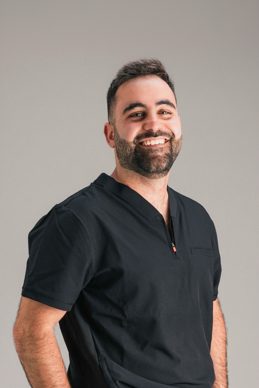
Alejandro Estrada Martínez centra su práctica en Rehabilitación Oral y Estética Dental. Ejerce en Álava. Se formó como odontólogo en Universidad del País Vasco en 2016.
Su recorrido formativo explica el resto: ha orientado su formación de posgrado hacia implantología y cirugía oral. Su título de posgrado más reciente es el Máster de Formación permanente en Rehabilitación protésica convencional e implantosoportada y transtornos témporo-mandibulares (Universidad de Oviedo, 2018).
Actualmente ejerce como prostodoncista en Clínica dental Reyes de Navarra (Álava, España).
Compagina la clínica con la dirección docente en EDE Posgrados y Universidad Europea Miguel de Cervantes (UEMC), en programas como Máster en Estética y Rehabilitación Oral y Experto en Prótesis sobre Implantes y Oclusión.
Dentro de Doctor&Cols firma 13 publicaciones científicas en revistas como Journal of Functional Biomaterials y Journal of Clinical Medicine.
Explora su trayectoria
Máster de Formación permanente en Rehabilitación protésica convencional e implantosoportada y transtornos témporo-mandibulares
Máster · Universidad de Oviedo · Oviedo, España
Especialización Universitaria Implantología Oral y Microcirugía
Especialista · Universidad del País Vasco · Leioa, España
Grado / Licenciado en Odontología
Grado · Universidad del País Vasco · Leioa, España
Clínica dental Reyes de Navarra · Álava, España
Director · Máster en Estética y Rehabilitación Oral
EDE Posgrados · Madrid, España
Co-Director · Experto en Prótesis sobre Implantes y Oclusión
EDE Posgrados · Madrid, España
Profesor colaborador · Máster de Formación Permanente Internacional en Cirugía Plástica Periodontal e Implantología
Universidad Europea Miguel de Cervantes (UEMC) · Valladolid, España
Journal of Functional Biomaterials
★ Alto impacto · Q1 · IF 5.16Autores · Esteban Pérez-Pevida, Iván Monteagudo-Villalobos, David Chávarri-Prado, Alejandro Estrada-Martínez, Miguel Beltrán-Guijarro, Markel Diéguez-Pereira, Aritza Brizuela-Velasco
Coautores Doctor&Cols · Markel Diéguez Pereira, Esteban Pérez Pevida, David Chávarri Prado, Miguel Beltrán Guijarro, Iván Monteagudo
Resonance frequency analysis (RFA) has been used as a diagnostic method to measure implant stability at all stages of healing. In addition to evaluating the status of the peri-implant marginal bone, it can also indicate the most appropriate time to load the implant. This in vitro study aimed to evaluate the efficacy of RFA as a diagnostic method for the detection of peri-implant marginal bone loss (MBL). Forty bone-level Klockner Vega implants were placed in a polyurethane block with elastic properties similar to those of the maxillary bone. The insertion torque and primary implant stability at the time of placement were measured using an RFA device. A circumferential peri-implant defect was created by removing the cortical bone portion in each implant using a trephine. The stability values were measured again using RFA. The stability values measured using RFA were lower after the creation of the circumferential peri-implant defect, indicating a statistically significant decrease in implant stability. The results of the study tend to show a relationship between peri-implant marginal bone loss and modifications in implant stability measured by RFA.
Journal of Clinical Medicine
★ Alto impacto · Q1 · IF 3.408Autores · Oier Montalbán-Vadillo, Esteban Pérez-Pevida, Iratxe Viteri-Agustín, David Chávarri-Prado, Alejandro Estrada-Martínez, Markel Diéguez-Pereira, Fernando Sánchez-Lasheras, Aritza Brizuela-Velasco
Coautores Doctor&Cols · Esteban Pérez Pevida, Markel Diéguez Pereira, David Chávarri Prado
Background: Although xenografts have shown successful results in GBR procedures due to their osteoconductive properties, many authors have opted to add co-adjuvant drugs to favor osteogenesis and differentiate cells into an osteoblastic lineage. Metformin has been shown to have bone-protective properties, regulating osteoclast differentiation, as well as the ability to promote osteoblast mineralization and differentiation. The present study aimed to evaluate the effect of the local application of a 1% metformin solution on bone neoformation in the treatment of an experimental bone defect in a guided bone regeneration animal model with a particulated bovine hydroxyapatite xenograft with hyaluronate. Methods: With this purpose in mind, two critical defects with 8 mm diameter and 0.5 mm depth were created in eight male New Zealand rabbit calvarias. Titanium cylinders were fixed in each defect and filled with particulate hydroxyapatite of bovine origin and sodium hyaluronate, with sterile injectable saline added to the control group and sterile 1% metformin solution added to the test group. At 6 weeks, the animals were euthanized, and samples were obtained and prepared for histomorphometric analysis. Results: A higher percentage of new bone formation was observed in the metformin samples than in the control samples, both in the region closest to the animal's calvaria and in the most distal region analyzed. A higher average bone–biomaterial contact percentage was observed in the samples, with metformin in both the proximal and distal regions. There was no statistically significant difference in the mean value in either region in both parameters. Conclusion: The local application of a 1% metformin solution in an animal model of guided bone regeneration with particulate bovine hydroxyapatite and hyaluronate resulted in greater bone neoformation and xenograft osseointegration than in the control group.
Journal of Functional Biomaterials
★ Alto impacto · Q1 · IF 5.284Autores · Miguel Beltrán-Guijarro, Esteban Pérez-Pevida, David Chávarri‐Prado, Alejandro Estrada-Martínez, Markel Diéguez-Pereira, Fernando Sánchez Lasheras, Aritza Brizuela‐Velasco
Coautores Doctor&Cols · Miguel Beltrán Guijarro, Markel Diéguez Pereira, Esteban Pérez Pevida, David Chávarri Prado
This study aims to analyse, using a finite element analysis, the effects of Ti-base abutment height on the distribution and magnitude of transferred load and the resulting bone microstrain in the bone-implant system. A three-dimensional bone model of the mandibular premolar section was created with an implant placed in a juxta-osseous position. Three prosthetic models were designed: a 1 mm-high titanium-base (Ti-base) abutment with an 8 mm-high cemented monolithic zirconia crown was designed for model A, a 2 mm-high Ti-base abutment with a 7 mm-high crown for model B, and a 3 mm-high abutment with a 6 mm-high crown for model C. A static load of 150 N was applied to the central fossa at a six-degree angle with respect to the axial axis of the implant to evaluate the magnitude and distribution of load transfer and microstrain. The results showed a trend towards a direct linear association between the increase in the height of the Ti-base abutments and the increase in the transferred stress and the resulting microstrain to both the prosthetic elements and the bone/implant system. An increase in transferred stress and deformation of all elements of the system, within physiological ranges, was observed as the size of the Ti-base abutment increased.
The International Journal of Periodontics & Restorative Dentistry
Q3 · IF 1.103Autores · David Chávarri-Prado, Andoni Jones, Esteban Pérez-Pevida, Markel Diéguez-Pereira, Alejandro Estrada-Martínez, Rodrigo Cabezón-Palacios
Coautores Doctor&Cols · Andoni Jones, Esteban Pérez Pevida, Markel Diéguez Pereira, David Chávarri Prado
Sinus floor augmentation is one of the most common approaches to obtain sufficient bone availability for placing implants in cases with severe bone atrophy in the posterior maxilla. Several bone substitutes are indicated for sinus augmentation, but they may achieve different clinical outcomes. This study aims to compare bovine bone mineral (BBM) with freeze-dried bone allograft (FDBA) in a two-stage lateral window sinus grafting approach. Twenty patients received a lateral window sinus elevation with either FDBA or BBM. Postoperative graft height was measured with CBCT. Implants were placed 6 months later, at which time biopsy samples were taken for histologic analysis and new CBCT scans were performed to measure graft height. The mean height reduction at 6 months was 20.27% ± 4.94% for FDBA samples and 5.36% ± 2.41% for BBM samples. The histologic analysis revealed a mean ratio of newly formed bone of 43.70% ± 5.29% for the FDBA group and 38.11% ± 4.03% for the BBM group. The FDBA group also showed a higher amount of residual biomaterial (17.25% ± 10.10%) and connective tissue (14.63% ± 4.38%) compared to the BBM group (15.53% ± 5.42% and 13.11% ± 4.42%, respectively). The differences between groups were statistically significant for the height reduction and newly formed bone (P ≤ .05) but not for the amounts of residual biomaterial and nonmineralized connective tissue (P ≥ .05). Six months after performing a lateral window sinus elevation, the percentage of newly formed bone was significantly higher when using FDBA than when using BBM, although the graft height reduction was also significantly higher for the FDBA group.
The International Journal of Oral & Maxillofacial Implants
Q2 · IF 1.088Autores · Chávarri-Prado D, Brizuela-Velasco A, Diéguez-Pereira M, Chento-Valiente Y, Estrada-Martínez A, Pérez-Pevida E, Álvarez-Arenal Á
Coautores Doctor&Cols · Esteban Pérez Pevida, David Chávarri Prado, Markel Diéguez Pereira
Purpose: The aim of this study was to measure and compare the retention degrees of different attachment systems before and after 1 year of mastication and removal/insertion simulations. Materials and methods: A 4 × 10-mm implant embedded in self-curing acrylic resin was placed inside a rectangular metal frame installed in a plastic container with artificial saliva at 37°C. Three different attachment systems were connected to the implants: Dalbo Classic, Locator (with blue, pink, and clear inserts), and Klockner Rotula. A loading test at 100 N and 1.25 Hz frequency was repeated 10 times for each attachment (changing the matrix component or plastic insert) until a total of 300,000 cycles and 1,095 removal/insertion sequences. The retention force was assessed at baseline until the end of the test (1 year) after 3-month intervals of simulation. One-way analysis of variance (ANOVA) and post hoc Tukey test (P < .05) were performed for statistical analyses. Results: Klockner Rotula showed the highest retention values at the beginning (69.6 ± 9.2 N; 7.1 ± 0.9 kg) and the end of the test (51.4 ± 5.4 N; 5.2 ± 0.6 kg), while Dalbo Classic showed the lowest values both at the beginning (13.1 ± 1.7 N; 1.3 ± 0.2 kg) and the end of the test (6.7 ± 1.6 N; 0.7 ± 0.2 kg). Locator with clear insert exhibited the lowest retention loss ratio (20.4%), while Locator with blue insert showed the highest (50.8%). At the end of the test, all attachment systems showed a significant decrease in retention force in comparison with the initial retention (P < .001). Conclusion: Locator attachments with clear and pink inserts and the Klockner Rotula attachment system showed retention forces > 20 N (2 kg) after 1 year of mastication and prosthesis insertion/removal simulation. All attachment systems showed a progressive loss of retention, which was higher for Locator attachments with blue and pink inserts and Dalbo Classic. The Klockner Rotula attachment system showed the highest retention values both at the beginning and the end of the test.
Coatings
★ Alto impacto · Q1 · IF 3.758Autores · Juan Carlos Vara, Juan J. Delgado, Alejandro Estrada-Martínez, Esteban Pérez-Pevida, Aritza Brizuela‐Velasco, Begoña M. Bosch, Román A. Pérez, Javier Gil
Coautores Doctor&Cols · Esteban Pérez Pevida
The placement of bone–level dental implants can lead to the detachment of particles in the surrounding tissues due to friction with the cortical bone. In this study, 60 bone–level dental implants were placed with the same design: 30 made of commercially pure grade 4 titanium and 30 made of Ti6Al4V alloy. These implants were placed in cow ribs following the company’s placement protocols. Particles detached from the dental implants were isolated and their size and specific surface area were characterized. The irregular morphology was observed by scanning electron microscopy. Ion release to the medium was determined at different immersion times in physiological medium. Cytocompatibility studies were performed with fibroblastic and osteoblastic cells. Gene expression and cytokine release were analysed to determine the action of inflammatory cells. Particle sizes of around 15 μM were obtained in both cases. The Ti6Al4V alloy particles showed significant levels of vanadium ion release and the cytocompatibility of these particles is lower than that of commercially pure titanium. Ti6Al4V alloy presents higher levels of inflammation markers (TNFα and Il–1β) compared to that of only titanium. Therefore, there is a trend that with the alloy there is a greater toxicity and a greater pro-inflammatory response.
BioMed Research International
Q3 · IF 1.081Autores · Esteban Pérez-Pevida, David Chávarri‐Prado, Markel Diéguez-Pereira, Alejandro Estrada-Martínez, Oier Montalbán-Vadillo, Antonio Jiménez-Garrudo
Coautores Doctor&Cols · Markel Diéguez Pereira, Esteban Pérez Pevida, David Chávarri Prado
Background and Objective . Marginal bone loss around dental implants is one of the most prevalent complication, and its biomechanical impact may be critical for treatment prognosis. The objective of this study was to evaluate the influence of marginal bone loss around dental implants in the occlusal load transfer to the bone in terms of magnitude of stress and strain and distribution of such transferred stress. Methods . Three models of a single internal connection bone level‐type implant inserted into a posterior mandible bone section were constructed using a 3D finite element software: one control model without marginal bone loss and two test models, both with a circumferential peri‐implant bone defect, one with a 3 mm high defect and the other one 6 mm high. A 150 N static load was tested on the central fossa at 6° relative to the axial axis of the implant. Results. The results showed differences in the magnitude of strain and stress transferred to the bone between models, being the higher strain found in the trabecular bone around the implant with greater marginal bone loss. Stress distribution differed between models, being concentrated at the cortical bone in the control model and at the trabecular bone in the test models. Conclusion . Marginal bone loss around dental implants under occlusal loading influences the magnitude and distribution of the stress transferred and the deformation of peri‐implant bone, being higher as the bone loss increases.
Journal of Oral Implantology
Q3 · IF 0.717Autores · Andoni Jones, David Chávarri‐Prado, Markel Diéguez-Pereira, Alejandro Estrada-Martínez, Miguel Beltrán-Guijarro, Esteban Pérez-Pevida
Coautores Doctor&Cols · Miguel Beltrán Guijarro, Markel Diéguez Pereira, Esteban Pérez Pevida, David Chávarri Prado
The purpose of this study was to determine the prevalence of favorable anatomy for palatal emergence of an immediate flapless implant in the maxillary central incisor post-extraction site. Implants were virtually placed into maxillary central incisor sites using 3-dimensional implant planning software. Following a strict implant placement criterion to keep a safe distance to the buccal plate and other anatomical structures, sockets were assessed to determine their suitability for an implant emerging from the palatal aspect. From 321 patients included in this study, 62.3% presented a suitable socket anatomy for an immediate implant to be placed with an angulation suitable for a screw-retained crown. In 29% of the cases, the implants had to be labially tilted to maintain a minimum distance to the buccal plate; 8.7% were unsuitable for immediate implants due to anatomic limitations. The position and angulation for an immediate implant in the maxillary central incisor socket should be carefully assessed preoperatively with 3-dimensional images, as many sites will not be candidates for a palatal emergence and thus a screw-retained restoration.
International Journal of Odontostomatology
Q4 · IF 0.197Autores · David Chávarri-Prado, Aritza Brizuela-Velasco, Pablo Ortiz-de-Urbina-Comerón, Markel Diéguez-Pereira, Alejandro Estrada-Martínez, Esteban Pérez-Pevida
Coautores Doctor&Cols · Esteban Pérez Pevida, David Chávarri Prado, Markel Diéguez Pereira
La estabilidad primaria es un requisito importante para la supervivencia y éxito de los implantes durante la osteointegración. En los últimos años, los implantes inmediatos postextracción han demostrado ser una opción de tratamiento exitosa y predecible para la reposición de dientes con mal pronóstico, pero surge la duda de si dichos implantes alcanzan valores de estabilidad primaria comparables a aquellos colocados en hueso maduro. Comparar la estabilidad primaria de implantes inmediatos colocados en alveolos postextracción con la de implantes colocados en hueso maduro. Se llevó a cabo un estudio clínico retrospectivo, con los datos recogidos sobre 175 implantes, colocados en 175 pacientes. Todos los implantes colocados pertenecían al modelo Essential Cone (Klockner Implant System) y se dividieron en dos grupos: implantes inmediatos (Grupo A, n=31) e implantes colocados en hueso maduro (Grupo B, n=144). La estabilidad primaria de todos los implantes se midió mediante torque de inserción y análisis de frecuencia de resonancia con Osstell ISQ. No se encontraron diferencias estadísticamente significativas respecto a la estabilidad medida a través del torque de inserción (26,29+10,07 Vs 25,76+9,72 N/cm) pero sí que se encontraron diferencias significativas en la medida de la estabilidad primaria mediante AFR, siendo inferiores los valores correspondientes a los implantes colocados en los alveolos post exodoncia (60,74 ± 6,17 en sentido VL y 62,19 ± 7.64 en sentido MD frente a 68,34 ± 6.26 en sentido VL y 69,29 ± 7.98 en sentido MD obtenidos en los implantes colocados en hueso maduro). El torque de inserción de los implantes inmediatos es similar al de los implantes colocados en hueso maduro, pero sus valores ISQ son significativamente inferiores, lo que demuestra un mayor grado de micromovimiento, y por consiguiente, un mayor riesgo de fracaso durante el período de osteointegración.
Journal of Clinical and Experimental Dentistry
Q3 · IF 1.368Autores · David Chávarri‐Prado, Aritza Brizuela‐Velasco, Markel Diéguez-Pereira, Esteban Pérez-Pevida, Antonio Jiménez-Garrudo, Iratxe Viteri-Agustín, Alejandro Estrada-Martínez, Oier Montalbán-Vadillo
Coautores Doctor&Cols · Markel Diéguez Pereira, Esteban Pérez Pevida, David Chávarri Prado
Background: This study aimed to evaluate the effect of the implant design and the presence of cortical bone in the primary stability, as well as analyze the differences between the stability measurements obtained by two different resonance frequency analysis (RFA) devices. Material and Methods: A total of 80 Klockner implants of two different models [40 Essential Cone implants (group A) and 40 Vega implants (group B)] were used. The implants were placed in two polyurethane blocks that simulated the mechanical properties of the maxillary bone. One block featured a layer of cortical bone that was absent from the other block. The primary stability of all implants was measured by insertion torque and RFA using two different devices: Penguin RFA and Osstell IDX. Results: Primary stability was superior in the cortical bone in both torque and RFA. In the block containing cortical bone, group A implants obtained a greater insertion torque than did group B. The insertion torque was lesser in the bone lacking cortex. Regarding the ISQ of the implants, group A presented higher values in the block with cortical bone, but the values were lower in the block without cortical bone. There were no significant differences between the values obtained from the Osstell IDX and Penguin RFA. Conclusions: The presence of cortical bone positively influences the primary stability of dental implants. The design of the implant also has a statistically significant influence on implant primary stability, although the impact depends on whether there is coronal cerclage or not. There were no statistically significant differences in the implant stability measurements obtained by two different devices.
Biology
★ Alto impacto · Q1 · IF 3.37Autores · David Chávarri‐Prado, Aritza Brizuela‐Velasco, Ángel Álvarez‐Arenal, Markel Diéguez-Pereira, Esteban Pérez-Pevida, Iratxe Viteri-Agustín, Alejandro Estrada-Martínez
Coautores Doctor&Cols · Markel Diéguez Pereira, Esteban Pérez Pevida, David Chávarri Prado
Objectives: To determine the effect of mechanical loading of bone on the stability and histomorphometric variables of the osseointegration of dental implants using an experimental test in an animal model. Materials and Methods: A total of 4 human implants were placed in both tibiae of 10 New Zealand rabbits (n = 40). A 6-week osseointegration was considered, and the rabbits were randomly assigned to two groups: Group A (Test group) included 5 rabbits that ran on a treadmill for 20 min daily during the osseointegration period; Group B (Controls) included the other 5 that were housed conventionally. The monitored variables were related to the primary and secondary stability of the dental implants (implant stability quotient—ISQ), vertical bone growth, bone to implant contact (BIC), area of regenerated bone and the percentage of immature matrix. Results: The results of the study show a greater vertical bone growth (Group A 1.26 ± 0.48 mm, Group B 0.32 ± 0.47 mm, p < 0.001), higher ISQ values (Group A 11.25 ± 6.10 ISQ, 15.73%; Group B 5.80 ± 5.97 ISQ, 7.99%, p = 0.006) and a higher BIC (Group A 19.37%, Group B 23.60%, p = 0.0058) for implants in the test group, with statistically significant differences. A higher percentage of immature bone matrix was observed for implants in the control group (20.68 ± 9.53) than those in the test group (15.38 ± 8.84) (p = 0.108). A larger area of regenerated bone was also observed for the test implants (Group A 280.50 ± 125.40 mm2, Group B 228.00 ± 141.40 mm2), but it was not statistically significant (p = 0.121). Conclusions: The mechanical loading of bone improves the stability and the histomorphometric variables of the osseointegration of dental implants.
BioMed Research International
Q3 · IF 1.081Autores · Markel Diéguez-Pereira, David Chávarri‐Prado, Alejandro Estrada-Martínez, Esteban Pérez-Pevida, Aritza Brizuela‐Velasco
Coautores Doctor&Cols · Markel Diéguez Pereira, Esteban Pérez Pevida, David Chávarri Prado
Objective . Long‐term clinical data on the success and complication rates of monolithic or minimally veneered zirconia implant‐supported restorations are lacking. Hence, the purpose of this retrospective clinical study was to analyze the complications of monolithic or partially veneered zirconia implant‐supported restorations up to 5 years follow‐up. Material and Methods . Single crowns, bridges, and full‐arch rehabilitations were included. The selection process was achieved by reviewing data from the prosthetic laboratory and excluding cases in which zirconium and full‐ceramic coating restorations were used. A total of 154 restorations were included (82 monolithic and 72 with buccal ceramic stratification). All the complications encountered, and the solutions applied, were explained. Results . A total of 93 restorative units had a follow‐up of between 24 and 60 months, and 61 restoration units had a follow‐up of between 12 and 24 months. A total of 7 complications were encountered (14.58% of cases; 95.45% per prosthetic unit). The technical complication rate was 2.08% (one case of minor chipping in one prosthetic unit); regarding the mechanical complications, four decementations (8.33% of the cases) and two screw loosening (4.17% of the cases) were encountered. Conclusions . Considering the limitations of this study, it can be concluded that monolithic or partially veneered zirconia implant‐supported restorations have a good clinical behavior during a follow‐up period of up to 5 years.
Journal of Dental Sciences
Q2 · IF 2.444Autores · Esteban Pérez-Pevida, Aritza Brizuela‐Velasco, David Chávarri‐Prado, Markel Diéguez-Pereira, Antonio Jiménez-Garrudo, Oier Montalbán-Vadillo, Iratxe Viteri-Agustín, Alejandro Estrada-Martínez, Gorka Santamaría-Arrieta
Coautores Doctor&Cols · Markel Diéguez Pereira, Esteban Pérez Pevida, David Chávarri Prado
BACKGROUND/PURPOSE: The use of osteotomes as a technique for densification and expansion of the residual ridge is one of the most widely used procedures to achieve adequate peri-implant bone quantity and density. The aim of this study was to evaluate the influence of time and the elastic deformation of peri-implant bone in the primary stability of implants placed using osteotomes. MATERIALS AND METHODS: In each of 10 fresh fragments of cow rib, two implants were placed using osteotomes. The insertion torque and initial implant stability quotient (ISQ) values were measured. In the control implants, the immediate removal torque was measured, while in the test implants, after 15 min of placement, ISQ values were measured again, and the removal torque was measured. RESULTS: There were significant differences between the ISQ values and between the insertion torque and removal torque at 15 min. The ISQ values (perpendicular/parallel) increased between the initial moment (64.4 ± 9/70.3 ± 5.9) and 15 min (66/71.4 ± 6.4). The removal torque at 15 min (12.4 ± 5.8) was lower than the insertion torque (15.9 ± 5.9). Compression of the trabeculae in contact with the implant placed using osteotomes was observed, as well as a greater number of trabecular fractures in the implants placed using conventional drilling. CONCLUSION: There is an increase in ISQ values of dental implants placed using osteotomes after 15 min of placement.
Rehabilitación Oral y Estética Dental
A Coruña · España
Rehabilitación Oral y Estética Dental
Madrid · España
Rehabilitación Oral y Estética Dental
Jaén · España
Rehabilitación Oral y Estética Dental
Barcelona · España
Rehabilitación Oral y Estética Dental
Barcelona · España
Rehabilitación Oral y Estética Dental
Argentina