Rehabilitación Oral y Estética Dental
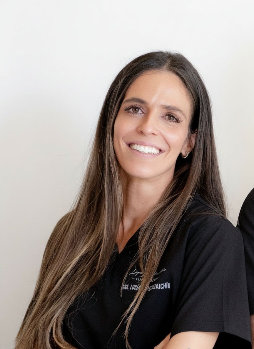
Lucía López Chaichío centra su práctica en Rehabilitación Oral y Estética Dental. Ejerce en Jaén. Se formó como odontóloga en Universidad de Granada en 2012.
Su recorrido formativo explica el resto: ha orientado su formación de posgrado hacia periodoncia, cirugía oral, regeneración, implantología y láser y ha llegado al nivel de doctorado. Su título de posgrado más reciente es el Doctorado en Odontología (Universidad de Granada, 2022). Ha pasado por Universidad de Granada y GC Formación.
Actualmente ejerce como director médico en Clínica Dental López Chaichío (Jaén, España).
Dentro de Doctor&Cols imparte 2 formaciones — entre ellas «Estancia clínica privada en cirugía mucogingival» y «Premium Clinical Mentoring en cirugía mucogingival» — en Clínica Dra. López Chaichío, Jaén y Clínica Dra. López Chaichío (Jaén) y clínica del alumno, ha publicado 7 casos clínicos sobre periodoncia, cirugía plástica periodontal y colgajo de avance coronal y firma 7 publicaciones científicas en revistas como Journal of Oral & Maxillofacial Research y Journal of Clinical Medicine.
Explora su trayectoria
Cirugía periodontal Dr. Andrea Tinti
Curso · SCOE · Barcelona, España
Monográfico de Regeneración Ósea Dr. Luca de Stavola
Curso · SCOE · Barcelona, España
Estancia Dr. Zucchelli Manejo de tejidos blandos alrededor de dientes e implantes (Bolonia)
Curso · Universidad de Bolonia · Bolonia, Italia
Curso de Estética Dental en Anteriores: El Arte de los Composites Anteriores
Curso · Autrán Dental Academy · Madrid, España
Doctorado en Odontología
Doctorado · Universidad de Granada · Granada, España
Máster Universitario en Periodoncia, Implantología Quirúrgica y Láser Odontológico
Máster · GC Formación · Alicante, España
Máster Universitario en Ciencias Odontológicas
Máster · Universidad de Granada · Granada, España
Experto en Cirugía y Rehabilitación Implantológica
Experto · Universidad de Sevilla · Sevilla, España
Grado / Licenciado en Odontología
Grado · Universidad de Granada · Granada, España
Clínica Dental López Chaichío · Jaén, España
Sin actividad docente registrada
 10 imgs
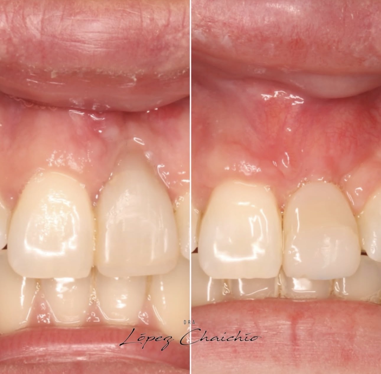
Paciente remitida por un defecto mucogingival en el 21 tras el fracaso de un implante inmediato. Sin opción de reimplantar, se resuelve la demanda estética con un colgajo de avance coronal con preservación de papila e injerto de tejido conectivo para compensar el déficit vertical y horizontal.
 10 imgs
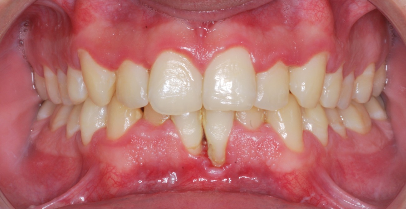
Problema periodontal severo en incisivos inferiores con ausencia de encía queratinizada apical al defecto, inserción marginal del frenillo y colapso de la papila 31-41. Se resuelve con la técnica del muro vestibular del Dr. Zucchelli: colgajo de avance coronal combinado con preservación papilar para acceder al defecto óseo, musculectomía, amelogeninas e injerto de tejido conectivo.
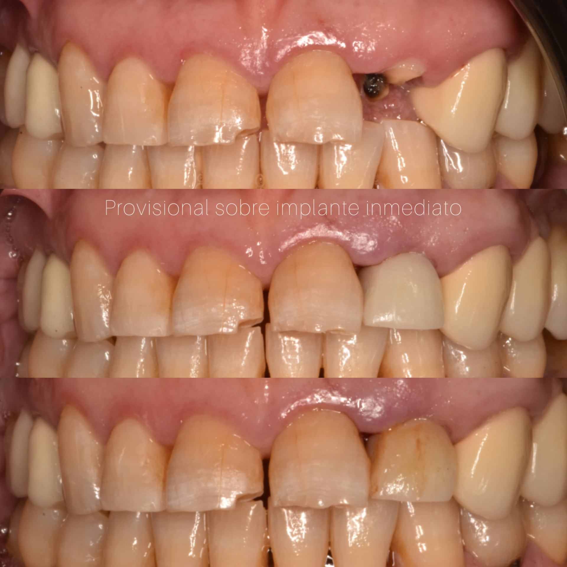
Caso de implante inmediato post-extracción en 2.2 con carga inmediata mediante provisional atornillado personalizado en clínica: preservación del perfil de emergencia, acondicionamiento gingival guiado por el provisional y transición a corona definitiva sin colapso del festón.
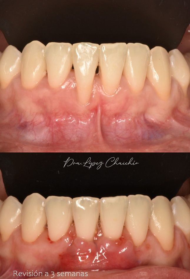
Cirugía mucogingival en el sector anteroinferior sobre biotipo muy fino con torque vestibular marcado e inserción alta de frenillos: musculectomía, injerto de tejido conectivo y colgajo de avance coronal, con control a tres semanas.
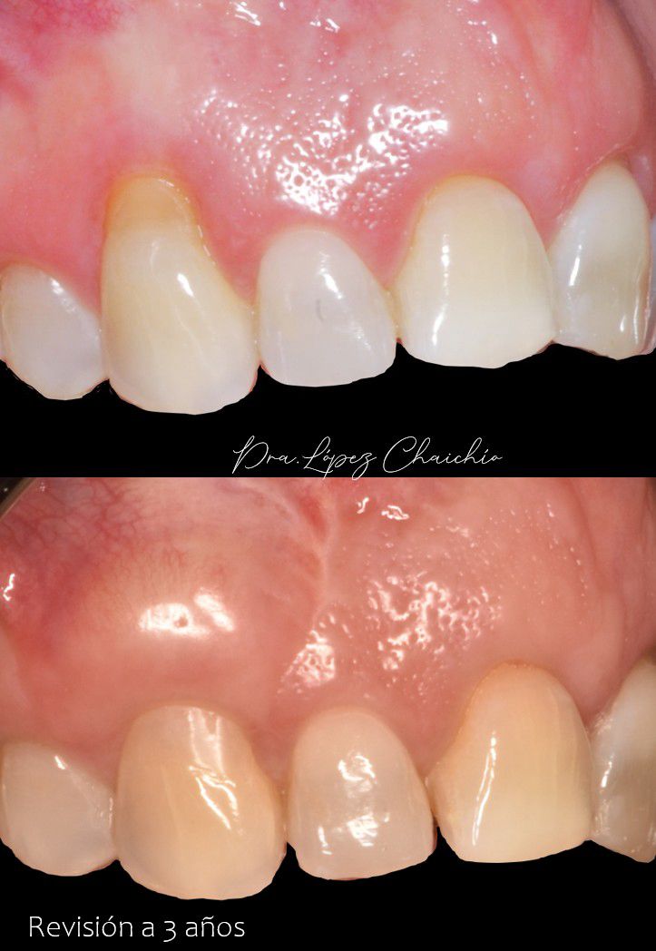
Recesión en el canino superior derecho tratada con injerto de tejido conectivo y colgajo de avance coronal, con control clínico a tres años que documenta la estabilidad del margen y del grosor de tejido.
 7 imgs
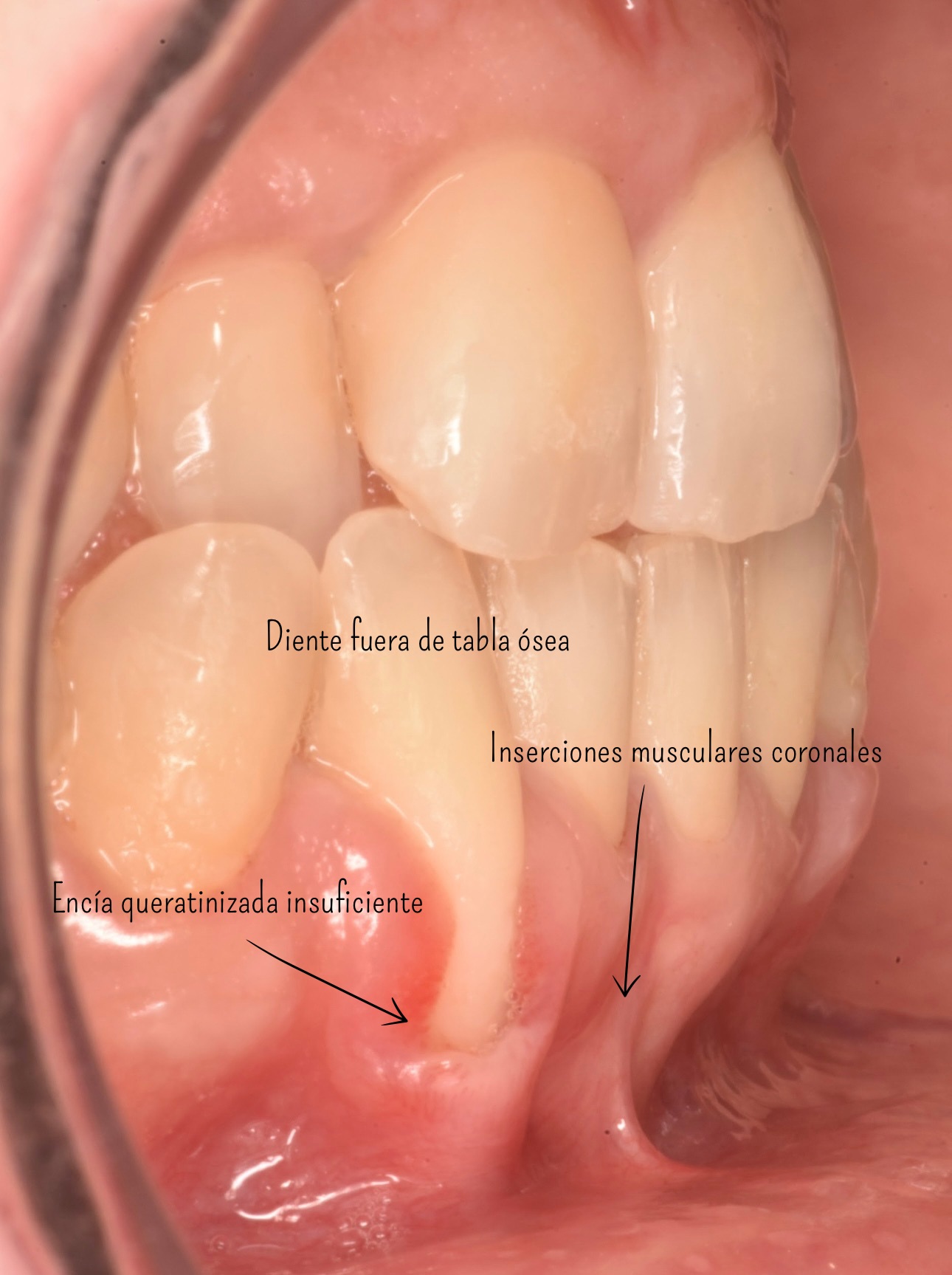
Recesión activa y dolor en un incisivo inferior fuera de tabla ósea, con encía queratinizada insuficiente e inserciones musculares coronales. Se decide un injerto de tejido conectivo previo al tratamiento de ortodoncia para asegurar la salud periodontal durante el movimiento dentario.
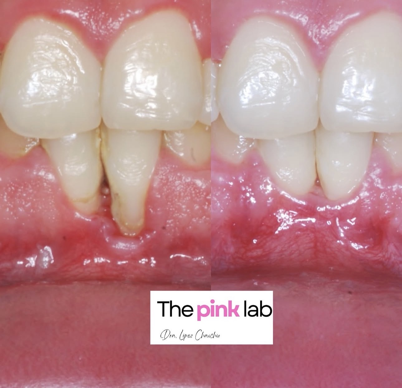
Recesión avanzada en incisivo inferior con pérdida de altura papilar y proximidad radicular extrema. Abordaje con la técnica del muro vestibular de Zucchelli e injerto de tejido conectivo, preservando y reconstruyendo la papila pese a la situación inicial desfavorable.
Curso 26-27 · Viernes por la mañana (9:30 - 14:00 h), fecha a consultar con la mentora · Clínica Dra. López Chaichío, Jaén · Presencial con mentoría on-line
Cirugía y Periodoncia · Cirugía Mucogingival
Curso 26-27 · Viernes por la mañana (9:30 - 14:00 h), fechas a consultar con la mentora · Clínica Dra. López Chaichío (Jaén) y clínica del alumno · Presencial con mentoría on-line
Cirugía y Periodoncia · Cirugía Mucogingival
Journal of Oral & Maxillofacial Research
Q2 · IF 1.439Autores · Lucía López-Chaichío, Pablo Galindo-Moreno, Miguel Padial-Molina, Lourdes Gutiérrez-Garrido, Roque Rodríguez-Álvarez, Francisco O'Valle, Andrés Catena
Objectives: The purpose of this observational clinical study was to evaluate the relationship between brain anatomical and volumetric changes in white matter, grey matter, and cerebral cortex thickness with the number of functional occlusal pairs present in the mouth. Material and Methods: The number of functional occlusal pairs in 70 patients was counted and non-invasive brain analysis was performed using magnetic resonance imaging. The volume of grey matter, white matter, and thickness of the cortex in different areas of the brain were determined by SPM12 and CAT12 software. Multiple regression model corrected for multiple comparisons using FDR and Spearman correlation coefficient were calculated for statistical comparison. Results: A total of 70 (39 male, 31 female) were analysed, with an average number of occlusal pairs of 10.21 (3.99). According to the Spearman correlation coefficient, a lower number of occlusal pairs was related to a reduction in white matter (right external capsule and posterior limb of the internal capsule), a reduction in grey matter (right temporal superior and medial gyrus and left cerebellum crus 1) and a reduction in thickness of the cerebral cortex (rostral anterior cingulated cortex of the right hemisphere and areas in the right and left hemisphere, especially in the frontal cortex). Conclusions: The number of occlusal pairs is related to the volume of white matter, grey matter, and thickness of the cerebral cortex in areas of the brain that are directly involved in the onset and progression of Alzheimer's disease and other dementias.
Journal of Clinical Medicine
★ Alto impacto · Q1 · IF 3.408Autores · Pablo Galindo-Moreno, Andrés Catena, Lucía López-Chaichío, Tiago Borges, Francisco O'Valle, Laura Torrecillas-Martínez, Miguel Padial-Molina
The aim of this study was to analyze the long-term marginal bone level (MBL) of implants supporting fixed full-arch restoration in patients who had previously lost their dentition due to severe periodontitis. This retrospective study included 35 patients in whom 342 implants with internal tapered conical connections were placed. MBL was analyzed radiographically over time and a long-term estimation of MBL was calculated. A mixed linear model with abutment height, graft, diameter and location (maxilla/mandible) as factors and gender, age, implant length and prosthetic variables as covariates was used to evaluate the influence on MBL. MBL in these patients showed an estimator of predictions at 4108 days after loading of −0.307 mm, SE = 0.042. Only 0.15% of implants were radiographically affected with MBL of 3 mm or more. The mixed linear model results showed a main effect of the type of opposing dentition, gender, implant diameter, and abutment height. Particularly, an abutment height of 1 mm had associated larger MBL than the remaining heights. Thus, it can be concluded that dental implants restored with fixed segmented full-arch rehabilitation in patients with a history of severe periodontal disease do not suffer important marginal bone loss if some specific factors are considered, mainly the use of long transmucosal abutments (≥2 mm).
Clinical Oral Investigations
★ Alto impacto · Q1 · IF 3.783Autores · Pablo Galindo-Moreno, Lucía López-Chaichío, Miguel Padial-Molina, Gustavo Avila-Ortiz, Francisco O'Valle, Andrea Ravidà, Andrés Catena
Objective: To investigate if there is epidemiological evidence of an association between edentulism and cognitive decline beside that currently available from limited sample-sized case series and cross-sectional studies considering limited co-variables. Materials and methods: Data from two USA national health surveys [NHIS 2014–2017 and NHANES 2005–2018] were analyzed using multinomial logistic regression to study the impact of type of edentulism and number of remaining teeth on memory and concentration problems. Age, gender, socioeconomic status, education level, cardiovascular health index, body mass index, exercise, alcohol, smoking habits, and anxiety and depression were used as covariates. Results: The combined population sample was 102,291 individuals. Age, socioeconomic status, educational level, anxiety and depression levels, and edentulism showed the highest odds ratios for cognitive decline. Number of teeth present in the mouth was found to be a predictor of cognitive status. This association showed a gradient effect, so that the lower the number of teeth, the greater the risk of exhibiting cognitive decline. Conclusions: Edentulism was found among the higher ORs for cognitive impairment. Clinical relevance: Maintenance of functional teeth through the promotion of oral health may contribute to the preservation of memory/concentration and other essential cognitive functions.
Journal of Clinical Medicine
★ Alto impacto · Q1 · IF 3.487Autores · Galindo-Moreno P, Concha-Jeronimo A, Lopez-Chaichio L, Rodriguez-Alvarez R, Sanchez-Fernandez E, Padial-Molina M
The aim of this study was to analyze the differences in terms of the marginal bone level (MBL) around implants with either an internal conical or an internal hexagonal implant–prosthesis connection. A randomized clinical trial included patients in need of a single implant-supported restoration. The implant–prosthesis connection was either internal conical or internal hexagonal while maintaining the same type of implant macro- and microarchitecture. Clinical and radiographical variables were registered up to 12 months of follow-up, including MBL. A total of 30 patients were included in the study. The main outcome variable, MBL 12 months after prosthesis delivery, was statistically different in both groups: −0.25 (0.12) vs. −0.70 (0.43) (conical vs. hexagonal; p = 0.033). Differences were also observed at the 3- and 6-month follow-up visits as well as for the MBL change from prosthesis delivery to the 12-month follow-up (−0.15 (0.13) vs. −0.56 (0.44); conical vs. hexagonal; p = 0.023). Correlations between MBL around the implants and radiographic measurements on the adjacent teeth, buccal bone to implant, tissue thickness or keratinized tissue were not significant neither globally nor when analyzed independently by group. In view of such results, it can be concluded that single-unit restorations with internal hexagonal-connection implants induce higher marginal bone loss after 12 months of follow-up from prosthesis delivery than internal conical-connection implants.
Gerodontology
Q3 · IF 1.435Autores · Lucia Lopez-Chaichio, Miguel Padial-Molina, Francisco O'Valle, Jose Antonio Gil-Montoya, Andres Catena, Pablo Galindo-Moreno
Introduction: Ageing leads to physiological cognitive decline that it is worsened in people with neurodegenerative diseases such as Alzheimer's disease. Despite the ongoing search for a solution to this cognitive decline, no effective remedies have been established. It has been determined that modifiable external factors, such as oral health and occlusal function, prevent cognitive decline. Objective: To analyse the primary interactions between occlusal function and cognitive functions. Main findings: Masticatory function is related to cognitive functions. In particular, current evidence, from both animal and human studies, suggests that the activation of masticatory muscles and proper mastication, with natural teeth or dental prosthesis, induces the release of several mediators and the activation of specific brain areas. Together, they result in higher neuronal activity, neurotrophic support, blood flow and the prevention of amyloid-beta plaque formation. Thus, all the components of the masticatory system must work together in order to preserve cognitive function. Conclusions: Available evidence suggests that oral and cognitive health are more interconnected than previously thought. Therefore, maintenance and adequate restoration of the whole masticatory system are important for the prevention of cognitive decline. In summary, oral and chewing health lead to healthy cognitive ageing.
Clinical Oral Implants Research
★ Alto impacto · Q1 · IF 3.588Autores · Pablo Galindo-Moreno, Miguel Padial-Molina, Lucia Lopez-Chaichio, Lourdes Gutiérrez-Garrido, Natividad Martín-Morales, Francisco O'Valle
Objectives To analyze a modified biphasic phycogenic biomaterial in comparison with anorganic bovine bone in maxillary sinus floor elevation in humans. Material and methods Eight male patients in need of bilateral two-stage sinus floor elevation were consecutively recruited for this randomized split-mouth study. A combination of autogenous cortical bone (ACB, 20%) and anorganic bovine bone (ABB, 80%) (ACB + ABB group) or ACB (20%) and modified biphasic phycogenic material (BP, 80%) (ACB + BP group) were randomly assigned to graft each sinus. Patients were followed up for 6 months post-surgery when bone samples were collected for analysis. Results Radiographically, bone height gain was statistically higher in the ACB + ABB versus the ACB + BP group. While the analysis of the biological compartments showed differences in non-mineralized tissue (39.15 ± 20.97% vs. 65.87 ± 28.59%, ACB + ABB vs. ACB + BP respectively; p = .018) and remnant biomaterial particles (22.62 ± 17.01% vs. 7.96 ± 8.57%, respectively; p = .028), the percentage of mineralized tissue (38.23 ± 17.55% vs. 24.14 ± 24.66%, respectively; p = .398) showed no statistically significant difference. In contrast, ACB + ABB biopsies showed higher Musashi-1-positive cells per mm2 compared to ACB + BP biopsies (811.49 ± 875.30 vs. 236.90 ± 280.81; p < .018), where the fusiform cells corresponded mainly with fibroblasts, as demonstrated by ultrastructural analysis. Conclusion Both combinations of materials exhibited bone formation after 6 months of healing in the maxillary sinus cavity. However, the combination with biphasic phycogenic biomaterial induced a higher radiographical vertical resorption and graft collapse in comparison with the combination with anorganic bovine bone, possibly due to a higher remodeling of the graft.
Clinical Oral Implants Research
★ Alto impacto · Q1 · IF 3.588Autores · Pablo Galindo-Moreno, Lourdes Gutierrez-Garrido, Lucia Lopez-Chaichio, Claudia Guerra-Lorenzo, Roque Rodriguez-Alvarez, Miguel Padial-Molina
Objectives To compare the marginal bone level around implants with a thin multi-phosphonate coated surface after either an early or conventional loading protocol. Material and methods A randomized pilot clinical trial was conducted. Dental impressions were obtained after either 4 (test) or 8 weeks (control) and single crowns screwed-in 2 weeks later. Several variables were evaluated including radiographical marginal bone level (MBL), patient's level variables, and those related to the restoration and surrounding tissues. These data were obtained at several time points up to a 1-year follow-up. Results Thirty-four patients were included in the study, 18 assigned to the test group. No differences at implant placement were detected for tissue thickness, keratinized mucosa, nor any other clinical or radiological variable. At the time of impressions, tissue was thinner in the test group (2.30 (0.46) versus 2.78 (0.66) mm, test versus control, respectively; p = .012) so shorter abutments were used in this group. Regardless, no significant changes in marginal bone level were detected neither within group along time nor between groups. The average MBL at the 1-year follow-up was −0.15 (0.32) versus −0.22 (0.37) (p = .443) (test versus control, respectively). None of the clinical or radiological variables evaluated had a determinant influence on the MBL at any visit nor group. Conclusion The use of implants with a multi-phosphonate coated surface for early loading offers successful radiographical outcomes 1 year after loading. MBL over time was not affected by taking the impressions 4 or 8 weeks after implant placement and loading them 2 weeks later.
Rehabilitación Oral y Estética Dental
Madrid · España
Rehabilitación Oral y Estética Dental
Guanajuato · México
Rehabilitación Oral y Estética Dental
México
Rehabilitación Oral y Estética Dental
A Coruña · España
Rehabilitación Oral y Estética Dental
Barcelona · España
Rehabilitación Oral y Estética Dental
A Coruña · España