Cirugía Oral e Implantología
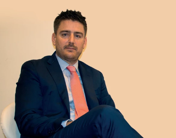
Juan Mesquida Escalas centra su práctica en Cirugía Oral e Implantología. Ejerce en Islas Baleares. Cursó sus estudios de Odontología en Loma Linda University (Estados Unidos) en 2008.
Su práctica diaria se desarrolla en Clínica dental Altes & Mesquida, como director médico (Islas Baleares, España).
Además de la clínica, mantiene actividad de docencia en Loma Linda University, en programas como Certificate in Implant Dentistry.
Su aportación al colectivo: ha publicado un caso clínico sobre implantología, cirugía oral y estética dental y firma 14 publicaciones científicas en revistas como The International Journal of Esthetic Dentistry y Frontiers in Dental Medicine.
Explora su trayectoria
Certificate in Implant Dentistry
Otro · Loma Linda University · Loma Linda, Estados Unidos
Internship in Implant Dentistry
Otro · Loma Linda University · Loma Linda, Estados Unidos
Clínica dental Altes & Mesquida · Islas Baleares, España
Profesor ayudante · Certificate in Implant Dentistry
Loma Linda University · Loma Linda, Estados Unidos
Autores · Juan Mesquida, Joseph Y. K. Kan, Herminio Garcia-Roncero, Matthew Fien, et al.
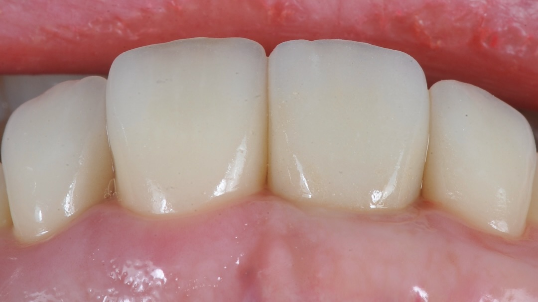
Este informe de dos casos analiza la estabilidad a largo plazo de las papilas interproximales alrededor de implantes dentales y revisa los aprendizajes acumulados durante las últimas dos décadas. A partir del seguimiento clínico y estético de pacientes rehabilitados con implantes, se exploran los factores anatómicos, protésicos y biológicos que pueden influir en la preservación o pérdida del tejido papilar. El trabajo ofrece una perspectiva clínica útil para la planificación implantoprotésica, el manejo de expectativas estéticas y el mantenimiento periodontal periimplantario.
The International Journal of Esthetic Dentistry
Q2 · IF 1.9Autores · Juan Mesquida, Diogo Firmino, Carmen Matji, Ignacio Ginebreda, et al.
Protocolo digital para reproducir el perfil de emergencia de una prótesis provisional de implante y transferir su macrogeometría a la restauración definitiva. Se combina un escaneo intraoral con el provisional in situ, un duplicado del escaneo para capturar la posición del implante y un escaneo extraoral de la prótesis, permitiendo fabricar una corona definitiva con adaptación óptima del tejido blando.
Autores · Matthew J. Fien, Israel Puterman, Juan Mesquida, Ignacio Ginebreda, et al.
This report presents two clinical cases exploring the use of fixation screws to stabilize membranes during guided bone regeneration, as an alternative to suturing the buccoapical periosteum. The approach aims to improve membrane positioning and stability while potentially simplifying management of augmented sites. By describing the clinical application of this technique, the article offers practical considerations for clinicians performing regenerative procedures and highlights an alternative strategy for membrane fixation in cases requiring predictable space maintenance and graft protection.
The International Journal of Esthetic Dentistry
Q2 · IF 1.9Autores · Juan Mesquida, Guillermo Bauza, Nadim Oliva, Ignacio Ginebreda, et al.
Background: Immediate implant placement (IIP) has shown predictable outcomes when specific requirements are met. The aim of the present study was to radiographically evaluate the dimensions of mandibular incisors and the alveolar bone to further determine the feasibility of IIP in this area. Materials and methods: A total of 404 intact mandibular incisor CBCT images from 101 patients (42 males, 59 females; mean age 49 ± 16.84 years) were analyzed. The mesiodistal tooth width and the distance between the incisal edge (IE), cementoenamel junction (CEJ), facial bone crest (BC), root apex (RA), and fenestration point (F) were registered, together with the alveolar bone width at 1 and 3 mm below BC. Finally, the angular discrepancy between the dental and the alveolar bone long axis (BLA) was recorded as the tooth torque (TT). Results: The CEJ to BC distance was 3.23 ± 1.67 mm. The RA to F mean distance was 15.02 ± 3.97 mm. The mean alveolar bone buccolingual widths at 1 and 3 mm below the facial BC were 7.12 ± 0.82 and 6.32 ± 0.71 mm in the lateral and central incisors, respectively (P 0.001). The buccolingual width was less than 6 mm in more than 33% of the central incisors, but only in 3% of the lateral incisors, displaying increased alveolar width. The average was 165.66 ± 7.47 degrees. Conclusions: The results presented in this study point toward the need for careful consideration of the requirements for anterior mandibular IIP for success and predictability purposes.
Frontiers in Dental Medicine
Q2 · IF 2.013Autores · Israel Puterman, Matthew J. Fien, Juan Mesquida, Ignacio Ginebreda, et al.
Regenerative medicine has gained much attention and has been a hot topic in all medical fields since its inception, and dentistry is no exception. However, innovations and developments in basic research are sometimes disconnected from daily clinical practice. This existing gap between basic research and clinical practice can only be addressed with improved communication between clinicians, academicians, industry, and researchers to facilitate the advance of evidence-based therapies and procedures and to direct research to areas of clinical need. In this perspective, six participants with strong clinical and research interests debated five previously conceived questions. These questions covered current methods and procedures for soft and hard tissue regeneration in the oral cavity with predictable outcomes, limitations of their respective protocols, and needs for future development of regenerative materials and technologies.
ResearchGate
Autores · Matthew Fien, Israel Puterman, Juan Mesquida, Guillermo Bauza, et al.
Stabilization of the graft-membrane complex during guided bone regeneration is a critically important aspect of implant dentistry. Several articles in the dental literature have introduced the utilization of periosteal biting stabilization sutures, rather than fixation screws and pins, to stabilize a bioabsorbable collagen membrane. This article reviews the concept of stabilization using sutures in periodontal regeneration and describes an alternative method, the Ribroast technique™, to stabilize a bioabsorbable membrane, whereby single periosteal biting horizontal mattress sutures are placed along the length of the defect to achieve sufficient stabilization. Two cases are presented that highlight the utility of this technique. Guidelines and limitations for the use of periosteal biting sutures are also discussed along with considerations and protocols that may be useful for improving treatment outcomes.
ResearchGate
Autores · Matthew J Fien, Israel Puterman, Juan Mesquida, Ferran Llansana Fitó, et al.
Several bioabsorbable membranes have been proposed to exclude soft-tissue ingrowth and to stabilize the bone graft when guided bone regeneration (GBR) is performed. The properties of the various membranes differ slightly due to variances in composition and manufacturing processes affecting their handling and suitability for specific techniques. The aim of this article is to present a technique to perform GBR with the use of a sugar-crosslinked absorbable collagen membrane in conjunction with a dehydrated amnion/chorion membrane (dHACM). This technique can be used to perform GBR at the time of tooth extraction and for ridge augmentation of an edentulous site in preparation for future dental implant placement. The use of a collagen membrane in combination with a dHACM can facilitate stabilization of the bone graft and membrane by providing the benefits of a long-lasting bioabsorbable collagen membrane in addition to the unique benefits of an amnion/chorion membrane, which has been shown to provide growth factors to the surgical site that have potential to enhance regenerative outcomes.
Journal of Prosthetic Dentistry
Q2 · IF 2.791Autores · Ferran Llansana, Igne Magne, Guillermo Bauza, Juan Mesquida
The biologically oriented preparation technique (BOPT) uses a vertical tooth preparation and simultaneously a controlled rotary instrumentation of the gingival sulcus. The resulting blood clot is stabilized with the interim restoration. However, accurate transfer of the emergence profile of the interim crown to the definitive restoration is essential to maintain the gingival architecture. This technique article describes clinical and laboratory procedures to predictably transfer the emergence profile of the interim crown to duplicate the gingival architecture in the definitive restoration.
The International Journal of Periodontics & Restorative Dentistry
Q3 · IF 1.136Autores · Israel Puterman, Matthew Fien, Juan Mesquida, Ferran Llansana
The aim of this report is to present a technique for buccal soft tissue contour augmentation with the use of a porcine volume-stable collagen matrix (VSCM). Augmentation of buccal soft tissue at the time of implant placement is often a necessity but is mostly performed using autogenous tissue. The technique using a VSCM can be done at the time of implant placement or, in the case of a two-stage procedure, at the time of implant uncovering. Here, clinical outcomes are reported in two cases when using VSCM concurrently with implant placement at sites in need of buccal contour augmentation to achieve a functional, esthetic result. The use of a xenograft poses several advantages over autogenous tissue while providing similar gains in soft tissue thickness. By eliminating the need to harvest a soft tissue graft from the palate, patient morbidity is reduced, and the reliance on palatal tissue thickness, to determine the amount of achievable augmentation, is eliminated.
Journal of Oral Implantology
Q3 · IF 0.717Autores · Cosimo Loperfido, Juan Mesquida, Jaime Lozada
A 71-year-old patient was successfully rehabilitated by means of a 3D-model-derived, hydroxyapatite-coated titanium subperiosteal mandibular implant. The implant was specifically designed to allow bone augmentation. The deficient bone was simultaneously grafted with mineralized bone allograft and recombinant bone morphogenetic protein-2 (rhBMP-2). The 32-month post-operative Cone Beam Computer Tomography (CBCT) follow-up showed vertical bone augmentation beneath the implant frame.
Elsevier eBooks
Autores · Nicholas Caplanis, Jaime Lozada, Juan Mesquida
Elsevier eBooks
Autores · Juan Mesquida, Jaime Lozada, Aladdin Al-Ardah, Sun Chun-xiao, Charles J. Goodacre
The International Journal of Oral & Maxillofacial Implants
Q3 · IF 1.071Autores · Phillip Roe; Joseph Y. K. Kan; Kitichai Rungcharassaeng; Joseph M. Caruso; Juan Mesquida; Jaime Lozada
This cone beam computed tomography study (CBCT) evaluated horizontal and vertical dimensional changes to the facial bone following maxillary anterior single immediate implant placement and provisionalization. CBCT scans taken immediately after (T1) and 1 year after surgery (T2) were evaluated. The midsagittal cut of each implant was identified, and measurements were made at predetermined levels. Horizontal facial bone thickness (HFBT) was measured at 0, 1, 2, 4, 6, 9, and 12 mm apical to the implant platform. Vertical facial bone level (VFBL) was the perpendicular distance from the implant platform (0) to the most coronal point of the facial bone. Measurements were recorded and changes between T1 and T2 were calculated. The data were analyzed statistically at a significance level of α = 0.05. CBCT scans of 21 patients were analyzed. At T2, the mean HFBT changes ranged from -1.23 to -0.08 mm at the seven different levels evaluated. The mean VFBL change was -0.82 mm. The HFBT changes at the 1- to 9-mm levels were not significantly different from one another, but they were significantly smaller than the change at the 0-mm level and significantly greater than the change at the 12-mm level. Significant positive correlations were observed only between horizontal and vertical changes and between horizontal change and initial VFBL at the implant platform. While the VFBL of eight implants (38%) was apical to the implant platform at T2, none was noted at T1. Dimensional changes to the peri-implant facial bone following maxillary anterior single implant placement and provisionalization should be expected. The greatest HFBT change was noted at the implant platform level, in part because HFBT change is correlated to the initial VFBL and the change in VFBL at that level.
Autores · Joseph Y. K. Kan, Kitichai Rungcharassaeng, Phillip Roe, Juan Mesquida, et al.
The purpose of this study was to determine the maxillary central incisor–incisive canal (CI-IC) relationship by comparing the visibility of the incisive canal on the two-dimensional sagittal images of the central incisors acquired from two cone beam computed tomography (CBCT) reconstruction modalities. A retrospective review of CBCT images from 60 patients (30 men and 30 women) with a mean age of 57.9 years (range: 19 to 83 years) was conducted. The CBCT images were evaluated using the manufacturer's recommended method (MRM) and a modified method (MM). The CI-IC relationship was classified as invisible, partially visible, or completely visible. The frequency distribution of the CI-IC relationships of the 60 samples indicated that the visibility of the incisive canal is significantly greater at all locations when using the MRM compared to the MM and on the right central incisor versus the left central incisor. Evaluation of the CI-IC relationship using the proposed method may allow the clinician to better determine whether additional surgical procedures are necessary when planning immediate implant placement and provisionalization at the maxillary central incisor positions.
Cirugía Oral e Implantología
Madrid · España
Cirugía Oral e Implantología
Barcelona · España
Cirugía Oral e Implantología
Madrid · España
Cirugía Oral e Implantología
Valencia · España
Cirugía Oral e Implantología
Madrid · España
Cirugía Oral e Implantología
Madrid · España