Cirugía Oral e Implantología
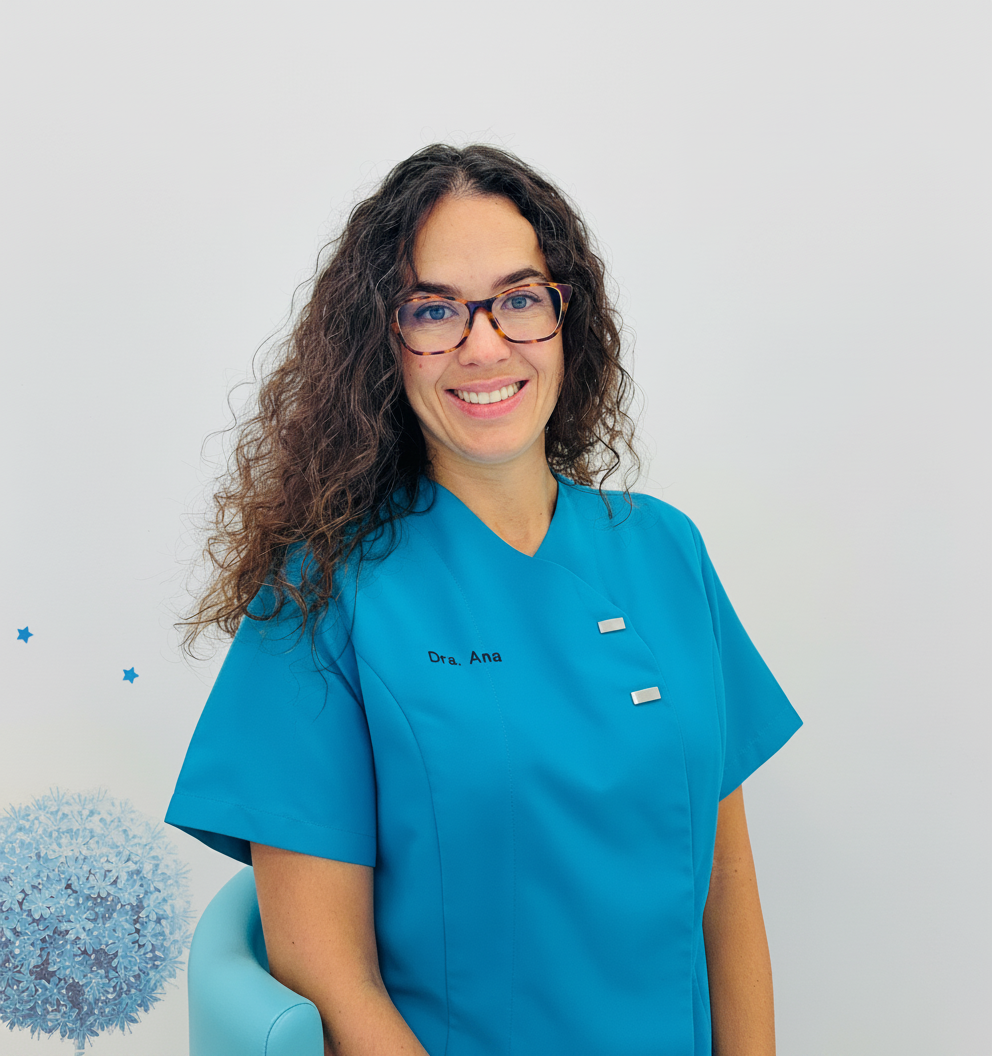
Ana Boquete Castro centra su práctica en Cirugía Oral e Implantología, con especial atención a cirugía guiada. Ejerce en Almería. Su formación de base procede de Universidad de Granada en 2008.
A lo largo de su trayectoria, ha llegado al nivel de doctorado y ha completado estudios de gestión clínica. Su título de posgrado más reciente es el Máster Universitario en Gestión y Planificación Sanitaria para Directivos de la Salud (Universidad Europea, 2023). Ha pasado por Universidad Europea, Universidad Católica San Antonio de Murcia (UCAM) y Universidad Internacional de Valencia.
Hoy trabaja como director médico en Clínica dental Integral - Dra. Ana Boquete (Almería, España). Antes desarrolló su actividad en Adeslas y Centro dental DEA.
La dirección docente ocupa la otra mitad de su tiempo: imparte clase en ACCA Learning, Universidad Alfonso X El Sabio y Posgrados Odontológicos PgO, en programas como Expertos Universitarios / Clínicos y Grado / Licenciado en Odontología.
En Doctor&Cols imparte 2 formaciones — entre ellas «Implantología Digital y Cirugía Guiada» y «Curso Universitario de Inmersión en Manejo de Bruxismo y Apnea Obstructiva del Sueño» — en Almería - España y firma 22 publicaciones científicas en revistas como Journal Of Clinical Periodontology y DOAJ (DOAJ: Directory of Open Access Journals).
Explora su trayectoria
Máster Universitario en Gestión y Planificación Sanitaria para Directivos de la Salud
Máster · Universidad Europea · Madrid, España
Máster en Formación del Profesorado
Máster · Universidad Católica San Antonio de Murcia (UCAM) · Murcia, España
Máster en Medicina Dental del Sueño
Máster · Universidad Católica San Antonio de Murcia (UCAM) · Murcia, España
Máster en Bioética Asistencial y Calidad y Seguridad en el Ámbito Sanitario
Máster · Universidad Internacional de Valencia · Valencia, España
Máster en Implantología Avanzada
Máster · Universidad de Murcia · Murcia, España
Doctorado en Ciencias Odontológicas
Doctorado · Universidad de Granada · Granada, España
Máster en Investigación Odontológica
Máster · Universidad de Granada · Granada, España
Diploma en Cirugía Bucal
Experto · Universidad Complutense de Madrid · Madrid, España
Máster en Implantología y biomateriales
Máster · Universidad de Murcia · Murcia, España
Grado / Licenciado en Odontología
Grado · Universidad de Granada · Granada, España
Clínica dental Integral - Dra. Ana Boquete · Almería, España
Adeslas · Madrid, España
Centro dental DEA · Almería, España
Asisa dental · Granada, España
Director · Expertos Universitarios / Clínicos
ACCA Learning · Barcelona, España
Profesor · Grado / Licenciado en Odontología
Universidad Alfonso X El Sabio · Madrid, España
Profesor · Máster Cirugía Oral, Implantes e Periodontologia
Posgrados Odontológicos PgO · Murcia, España
2ª edición: 16-17 octubre 2026 · 3ª edición: 20-21 noviembre 2026 · Almería - España · Presencial
Cirugía y Periodoncia · Implantología Digital
Fecha y sede por determinar
Armonización Orofacial
Journal Of Clinical Periodontology
★ Alto impacto · Q1 · IF 5.111Autores · Alexander De Greef, Kato Van Caesbroeck, Hanne Langers, Amy Vanrusselt, Pierre Lahoud, Ana Boquete‐Castro, Andy Temmerman
AIM: To assess how suture diameter, material composition and tissue characteristics influence the tension-to-tearing behaviour of oral soft tissues, with the objective of optimising atraumatic suturing. MATERIALS AND METHODS: This in vitro cadaver study evaluated 288 standardised porcine oral soft-tissue specimens (144 gingiva, 144 lining mucosa). Twelve different suture types including four distinct diameters (4-0, 5-0, 6-0, 7-0) and three different materials (polyvinylidene fluoride [PVF], polyamide [PA] and polyglycolic acid [PGA]) were tested using a calibrated Instron microtester. Maximum rupture force (N), failure mode and tissue thickness (mm) were recorded. Statistical analysis relied on three-way ANOVA, Tukey's post hoc testing and Pearson's correlation (α = 0.05). RESULTS: The overall mean maximum rupture force was 5.03 ± 3.00 N. Rupture force increased with suture diameter (p = 0.0001) and differed between materials (p < 0.0001; PGA ≈ PVF > PA). Gingival specimens resisted higher loads than mucosal specimens (p = 0.003). Fine sutures (6-0, 7-0) failed mainly by thread rupture, while thicker sutures (4-0, 5-0) caused tissue tearing, supporting the concept of a mechanical safety buffer. PA (6-0) generally preserved tissue integrity, whereas PVF and PGA required 7-0 in delicate mucosa to ensure comparable tissue protection. Tissue thickness correlated positively with rupture force (r = 0.49, p < 0.001). CONCLUSIONS: Finer sutures (6-0 and 7-0) function as protective mechanical fuses, safeguarding delicate tissues during manipulation. PA 6-0 provides reliable protection, while 7-0 PVF and PGA are recommended for fragile mucosa manipulation. These findings provide a biomechanical framework for evidence-based suture selection in periodontal plastic surgery.
DOAJ (DOAJ: Directory of Open Access Journals)
Autores · Kirenia Pieri Silva, Yordany Boza Mejías, Ana Boquete‐Castro
Fundamento: El quiste periapical es una lesión inflamatoria común de los maxilares; generalmente cursa de forma asintomática, puede llegar a al alcanzar un tamaño significativo y causar destrucción ósea; además en ocasiones desplazamiento de los dientes adyacentes. Objetivo: Mostrar el caso de un paciente que luego de la exéresis de un quiste periapical se le aplicó tratamiento de fibrina rica en plaquetas combinado con xenoinjerto logrando la conservación de los dientes. Reporte del caso: Presentamos el caso clínico de un paciente que acudió a consulta estomatológica, asintomático, con endodoncia fallida por sellado defectuoso en apical del diente número 31; al examen clínico se observó tejido blando sin soporte óseo a nivel de los dientes 31 y 41, al examen radiográfico se evidenció imagen radiolúcida hipodensa, bien definida, con ruptura de tabla vestibular y compromiso mucoso. La lesión fue tratada quirúrgicamente mediante exéresis del quiste periapical, apicectomía y regeneración ósea con xenoinjerto y fibrina rica en plaquetas (L-PRF). En el control posoperatorio al mes se observó adecuado proceso de cicatrización y ausencia de sintomatología. Después de un mes en el control posoperatorio, se observó adecuado proceso de cicatrización y ausencia de sintomatología. Conclusiones: Este caso muestra la efectividad de la terapia con plasma rico en fibrina combinado con xenoinjerto para el tratamiento de los defectos óseos producidos por un quiste periapical y la conservación de los dientes cercanos a la lesión.
Journal of Periodontal Research
★ Alto impacto · Q1 · IF 6.385Autores · Ana Boquete‐Castro, Jill Hadisurya, Mrinal Gaurav Srivastava, Kristel Bernaerts, Annabel Braem, Naiera Zayed
Multispecies oral biofilms were established on titanium implants using a controlled bioreactor model and subjected to different decontamination strategies. Biofilm persistence was evaluated by v-qPCR, SEM, and CLSM to assess treatment efficacy.
Bioengineering
★ Alto impacto · Q1 · IF 4.791Autores · Juan Algar, Cristian Docampo-Vázquez, Cristina Rico‐Romano, Ana Boquete‐Castro, C. Obispo, Juan Manuel Aragoneses
Apical periodontitis is a common inflammatory condition associated with root canal treatment (RCT) failure. The quality of the three-dimensional root canal seal is critical to the success of the treatment. Bioceramic sealants, such as Neosealer Flo, offer biological advantages such as osteoconduction, biocompatibility and sustained calcium ion release, which may improve apical healing. The aim of this study was to compare AH Plus and Neosealer Flo in terms of postoperative pain, extrusion and periapical healing. A single-blind, randomised clinical trial was conducted with 60 patients divided into AH Plus and Neosealer Flo groups. Post-operative pain was assessed using a visual analogue scale (VAS) at 24 and 48 h and at 7 days. Seal quality and periapical healing were assessed at 6 months using the AAE success criteria by clinical and radiographic evaluation. Neosealer Flo resulted in less postoperative pain at 24 h and 7 days compared to AH Plus. Extrusion did not significantly affect pain or correlate with the type of sealer used. Both materials achieved similar periapical healing rates. Neosealer Flo demonstrated advantages in pain reduction, while both sealants showed comparable efficacy.
The International Journal of Oral & Maxillofacial Implants
Q3 · IF 1.088Autores · Ricardo Medina Madrid, Estebon Padullés Roig, Guillermo Cabanes Gumbau, Raquel Alarcón, Ana Boquete‐Castro
PURPOSE: To determine the relationship between bone loss that occurs during the peri-implantitis process and variations in implant stability using resonance frequency analysis (RFA) measurement methods. MATERIALS AND METHODS: Forty selftapping implants were placed in cow ribs, and study scenarios were established according to the affected implant side and bone loss depth (n = 10 implants per group): Case 1 = bone loss on one side (vestibular); Case 2 = bone loss on two opposite sides (buccal and lingual); Case 3 = bone loss on two adjacent sides (buccal and mesial); and Case 4 = foursided bone loss (circumferential). For each group of 10 implants, first a bone loss of 0 mm was evaluated, then 4-mm defects (simulating 1/3 of bone loss) were created and evaluated, and finally 8-mm defects (simulating 2/3 of bone loss) were created and evaluated. Osteotomy measurements were made with a periodontal probe. For each implant, RFA was measured by the same operator using the Beacon system (Osstell). RESULTS: The initial implant stability quotient (ISQ) values of the 40 implants exceeded 70, reflecting an average of 73 in the buccolingual (VL) and 74.8 in the mesiodistal (MD) directions. ISQ measurements in the 10 implants in which bone dehiscence was performed on the vestibular aspect reflected a decrease in ISQ values as bone loss increased. When generating bone loss in two opposite sides (buccal and lingual), a greater decrease in ISQ values was observed when 2/3 of the implant were affected. The average VL ISQ measurement was less than 70 when at sites with 2/3 of bone loss. CONCLUSIONS: When bone loss occurs on only one side of the implant, the ISQ values decrease, but the implant maintains good stability. The same occurs when two opposite sides of the implant are affected, as the unaffected side has the least decrease in ISQ value.
Journal of Esthetic and Restorative Dentistry
★ Alto impacto · Q1 · IF 3.306Autores · Irene Maniega‐Mañes, Manuel Monterde‐Hernández, Karla Mora‐Barrios, Ana Boquete‐Castro
INTRODUCTION: AI is based on automated learning algorithms that use large bodies of information (big data). In the field of dentistry, AI allows the analysis of radiographs, intraoral images and other clinical recordings with unprecedented precision and speed. Facial analysis is known for helping dentists and patients achieve a satisfactory result when a restorative treatment must be realized. The objective of this study is to conduct a neural network-based computerized facial analysis using Python programming language in order to valuate its efficacy in facial point detection. METHODS: The neural network was trained to identify the main facial and dental points: smile line, lips, size and for of the teeth, etc. A facial analysis was carried out using AI. A descriptive analysis was made with calculation of the mean and standard deviation (SD) of the precision and accuracy in each group. Analysis of variance (ANOVA) was used for the comparison of means between groups. RESULTS: At the intersecting point between dentistry and technology, advances in artificial intelligence (AI) are producing a change in the way modern dentistry is performed. The present study evidenced lesser variability in the execution times of the neural network compared with the DSD system. This indicates that the neural network affords more consistent and predictable results, representing a significant advantage in terms of time and efficacy. CONCLUSION: The neural network is significantly more efficient and consistent in performing facial analyses than the conventional DSD system. The neural network reduces the time needed to complete the analysis and shows lesser variability in its execution times.
Sleep Science
Q4 · IF 0.529Autores · Juan-Manuel Cortes-Mejia, Ana Boquete‐Castro, Yoaly Arana-Lechuga, Guadalupe Terán-Pérez, Katiuska Casarez-Cruz, Rosa Obdulia González-Robles, Javier Velázquez‐Moctezuma
Objectives: This study aimed to evaluate the therapeutic effcacy of custom-made mandibular advancement devices (MAD) in the control of primary snoring and sleep apnea and to correlate with anatomical changes identified through imaging tests. Methods: Patients (n = 17) diagnosed with sleep apnea or primary snoring were included in this study and subsequently treated with MADs. Changes were assessed using a polysomnographic study (PSG), the Epworth Sleepiness Scale (ESS), and an imaging study with computed tomography scanning (CT). Studies were performed before and after the use of MAD. Anteroposterior measurements were taken in the sagittal plane at the hard palate, glottis, and supraglottic levels along the hard palate axis. Afterward, measurements were taken in the axial plane at the same levels along the hard palate axis. Results: From the six recorded measurements, the airway caliber increased by five. However, these changes were significant only in two measurements (sagittal hard palate and axial supraglottic). Snoring was controlled in 16 of the 17 subjects. From these sixteen, 12 subjects had a correct opening of the airway at the hard palate level. Moreover, daytime sleepiness decreased in all subjects. Discussion: Present results suggest that sagittal hard palate and axial supraglottic opening after use of MAD are mainly responsible for eliminating snoring and improve sleep apnea.
Revista Odontológica Mexicana Órgano Oficial de la Facultad de Odontología UNAM
Q4 · IF 0.075Autores · Juan Manuel Cortés-Mejía, Ana Boquete‐Castro
Introducción: las patologías cardiovasculares tienen elevada prevalencia. Asimismo, la prevalencia de trastornos respiratorios de sueño en pacientes con fallo cardiaco crónico es muy elevada. Objetivos: analizar el impacto del uso de dispositivos de avance mandibular (DAM) en pacientes con historia de fallo cardiaco previo. Material y métodos: se realizó una búsqueda bibliográfica electrónica con los términos MeSH occlusal splints, sleep apnea, obstructive y heart failure. Resultados: la severidad del trastorno respiratorio de sueño varía de acuerdo con la severidad de la patología cardiaca existente. Discusión: los efectos nocivos inducidos por la apnea obstructiva del sueño (AOS) en el sistema cardiovascular pueden implicar dos componentes: hipoxia intermitente crónica durante la apnea o hipopnea y grandes esfuerzos inspiratorios. Esto sobrecarga el miocardio alterando su estructura y función. El tratamiento con DAM reduce la gravedad de la AOS y aumenta la saturación de oxígeno en pacientes con fallo cardiaco congestivo. Además, al mantener las vías respiratorias superiores libres, el DAM reduce la presión intratorácica. Conclusiones: la prevención de complicaciones cardiacas en pacientes con AOS está relacionada con la prevención de eventos respiratorios.
Saudi Endodontic Journal
Q3 · IF 1.478Autores · Ana Boquete‐Castro, AlejandroPeña López, AlineSavariz Martins, AlbertoSierra Lorenzo, P Gonzalez Perez
Introduction: To assess the potential uses and benefits of cone-beam computed tomography (CBCT) in the field of endodontics, where the anatomy evaluated is complex, and to summarize the possible advantages over two-dimensional (2D) radiography. Methods: For this study, a review of the literature of the last 13 years was carried out in the PubMed and Google Scholar databases by using the following keywords: CBCT, dental radiography, endodontic diagnosis, management of endodontic problems; 3D imaging. Moreover, inclusion and exclusion criteria were applied. Results: A total of 29 articles were selected because they fulfilled the inclusion criteria. Articles were analyzed to investigate the advantages, limitations, radiological aspects, and uses of CBCT in endodontics from a critical point of view. Conclusions: Conventional radiography continues to be the main diagnostic method in endodontics, but taking into account the as low as reasonably achievable principle, in cases in which there are inconclusive signs and symptoms with 2D radiography, CBCT is a useful complement in endodontic diagnosis and allows a more accurate and predictable treatment plan design.
Journal of Clinical Dentistry and Research
Q4 · IF 0.037Autores · José Antonio Martín Hernández, Karla Daniela Mora, Ana Boquete‐Castro, Sidney Kina
Introdução: As resinas acrílicas têm sido clinicamente utilizadas por muitos anos em próteses dentárias. Avanços nos materiais CAD/CAM e desenvolvimentos recentes em nanotecnologia têm permitido o uso do grafeno como fase de reforço do poli (metacrilato de metila) (PMMA) para restaurações CAD/CAM, com resultados promissores. Material e Métodos: 96 amostras de discos monocromáticos G-CAM (G-MONO) e discos multicromáticos G-CAM (G-MULTI) foram igualmente divididas em quatro grupos (n=24): Grupo 1 (G-MONOc) e Grupo 2 (G-MULTIc) serviram como controle e não foram termociclados; o Grupo 3 (G-MONOt) e o Grupo 4 (G-MULTIt) foram submetidos à termociclagem. As medições foram feitas usando um durômetro digital Shore A, e os valores foram analisados estatisticamente usando ANOVA de um critério, seguida pelo teste de Tukey. Resultados: Os resultados foram muito semelhantes para G-MONO termociclado e não termociclado; a ANOVA de um critério não revelou diferenças significativas entre o Grupo 1 (G-MONOc) e o Grupo 3 (G-MONOt) e entre o Grupo 2 (G-MULTIc) e o Grupo 4 (G-MULTIt). Todas as comparações do teste de Tukey pareado também não apresentaram diferenças significativas (α<0,05). Conclusão: O PMMA para CAD/CAM modificado por grafeno passou, com sucesso, pelo processo de envelhecimento composto de 10.000 ciclos térmicos, mantendo seus valores de microdureza superficial. A manutenção da microdureza superficial propicia um menor desgaste e maior longevidade da restauração, assim permitindo a utilização do PMMA modificado por grafeno em restaurações permanentes. Outras investigações são necessárias para estabelecer as propriedades mecânicas do PMMA modificado por grafeno.
Journal of Clinical Dentistry and Research
Q4 · IF 0.037Autores · Jon Salazar, Ana Boquete‐Castro, Vicente Gimeno, Sidney Kina
Objetivo: O objetivo da presente revisão sistemática foi analisar se há diferenças com relação a preferência, satisfação e conforto dos pacientes, quando realizam, no consultório, moldagens convencionais ou digitais. Métodos: Foi realizada um pesquisa bibliográfica nas bases de dados Medline, Embase, Web of Science, Central e Clinical Trials, de acordo com a estratégia PICO. O risco de viés foi avaliado utilizando uma ferramenta da colaboração Cochrane, e o protocolo foi registrado no PROSPERO. Resultados: A busca sistemática inicial encontrou 79 artigos. Após a remoção dos duplicados e daqueles que não preenchiam os critérios de inclusão, restaram 10 estudos. Sete estudos incluídos demonstraram resultados mais favoráveis para a moldagem digital com relação ao conforto, satisfação e preferência; enquanto dois apresentaram resultados mais favoráveis para as moldagens convencionais. Um dos estudos não apresentou informações conclusivas entre a intervenção e a comparação. Foram realizadas duas metanálises de subgrupos com quatro artigos, de acordo com a marca do scanner e o tipo de randomização. Conclusões: houve alta heterogeneidade nos métodos utilizados nos ensaios clínicos randomizados (ECRs) disponíveis. A maioria não contribuiu com informação suficiente para analisar e integrar a estatística. Embora as conclusões devam ser interpretadas com cautela, a evidência científica atual sugere que a percepção dos pacientes com relação ao conforto, preferência e satisfação é favorável aos scanners intrabucais. É necessário realizar mais ECRs que incluam medidas do desfecho relatado pelo paciente.
Journal of oral medicine
Autores · Ana Boquete‐Castro, Lucero Sánchez Af
Background: Temporomandibular dysfunction (TMD), which is considered the main cause of non-dental orofacial pain. Therefore, treatment must be focused on the reduction of symptoms. In this sense, low intensity laser therapy is suggested to have bio stimulating and analgesic effects. Methods: A Cochrane and Medline databases search were conducted between June and December of 2017. The terms used for the different searches were “temporomandibular joint”, “laser therapy”, disorders, laser and treatment. Findings: 6 publications were selected from a total of 162. They were stratified using the SORT criteria (Strength of Recommendation Taxonomy). Conclusion: TMD is a complex disorder that can be challenging to diagnose and treat. The efficacy of laser in the treatment of TMD is rather promising. According to the literature reviewed, low level laser seems to have a benefit on patients with TMD, however, it is still difficult to establish a therapeutic protocol for TMD.
Materials
★ Alto impacto · Q1 · IF 4.287Autores · José Luís Calvo‐Guirado, Aldo Lucero-Sánchez, Ana Boquete‐Castro, Marcus Abboud, Sérgio Alexandre Gehrke, Manuel Fernández Domínguez, Rafael Delgado-Ruíz
The aim of this study was to evaluate the soft tissue thickness and marginal bone loss around dental implants with sloped micro-threaded shoulder (30° angle) in comparing with conventional design, inserted 30° degrees angulated in post extraction sockets and immediate loaded with temporary prosthesis simulating the all-on-four protocol. Materials and Methods: Six fox hound dogs received forty-eight post extraction dental implants with the different diameter and length (Medentika, Germany), but with different neck configurations. Two group of implants were inserted 1mm subcrestal. Control group has a micro-threaded neck and the Test group has a sloped microthreaded neck. Immediate loading was applied using a constructed metallic structure. After three months, soft and hard tissue levels were assessed by histomorphometric analysis. Results: The mean soft tissue thickness (STT) was 2.5 ± 0.2 mm for the Control group and 3.3 ± 0.3 mm for Test group (p = 0.036), meanwhile the mean marginal bone loss (MBL) was 1.53 ± 0.34 mm for Control group and, 1.62 ± 0.22 mm for Test group (p > 0.05). Conclusions: Within the limitations of this experimental model in dogs, the findings showed that dental implants with microthreaded and microthreaded sloped necks installed in immediate post extraction sites with immediate load, presented a comparable perimplant tissue behavior.
DOAJ (DOAJ: Directory of Open Access Journals)
Autores · José Luís Calvo‐Guirado, C. PÉREZ-ALBACETE, Consuelo Pérez Sánchez, Ana Boquete‐Castro, José Eduardo Maté Sánchez de Val, Joseph E. Peña, Maria Piedad Ramírez Fernández, Miguel Garcés, Luis Meseguer‐Olmo, Gerardo Gómez Moreno
Aim The purpose of this study was to determine if different melatonin concentrations enhances human adult mesenchymal stem cells (MSCs) differentiation into osteoblasts, in comparison with MSCs cultured with dexamethasone (DEX). Material and Methods MSCs were treated with different melatonin concentrations. Specifically: Group I: untreated MSCs; Group II: MSCs exposed to physiological doses of melatonin of 0.01 µM; Group III: MSCs exposed to 50 µM melatonin; Group IV: MSCs exposed to 100 µM melatonin; Group V: MSCs exposed to 150 µM melatonin; Group VI: MSCs exposed to 100 µM of DEX. Cell viability, adhesion, growth, differentiation and activity were evaluated at different time points (3, 7, 14, 21 and 28 days) using the following assays MTT, SEM, Flowcytometry, ALP activity, Alizarin Red staining and RT-PCR. Results Melatonin stimulated the viability and alizarin red activity of MSCs in a dose-dependent manner. Melatonin 50 µM significantly increased ALP activity, especially after 21 and 28 days of culture. A significant decrease in the expression of membrane markers CD75, CD105 and CD90 was recorded over time, in the presence of melatonin; therefore, the MSCs were early differentiated into osteoblasts regardless of the melatonin concentration. Conclusion These results demonstrated that melatonin directly accelerated the differentiation of human stem cells into osteoblasts and also suggested that melatonin could be applied as a pharmaceutical agent to promote bone regeneration.
Clinical Oral Implants Research
★ Alto impacto · Q1 · IF 3.681Autores · Ana Boquete‐Castro, Gerardo Gómez Moreno, José Luis Calvo‐Guirado, Antonio Aguilar‐Salvatierra, Rafael Delgado-Ruíz
OBJECTIVES: The aims of this meta-analysis were (i) to perform a systematic review of the relation between treatment with denosumab and the incidence of osteonecrosis of the jaw (ONJ) and (ii) to obtain information on dosage, first event apparition, and treatment approaches for patients with ONJ related to denosumab. MATERIALS AND METHODS: A systematic review and meta-analysis of relevant literature was performed in the PubMed, MEDLINE, Embase, and Cochrane databases, identifying randomized clinical trials that evaluate the adverse effects of denosumab. The overall incidence rates and 95% confidence intervals (CI) for ONJ were calculated employing fixed- and random-effects models, according to the heterogeneity of the studies included. RESULTS: A total of 8963 patients with a variety of solid tumors reported in seven randomized controlled trials (RCTs) were included in the systematic analysis. The overall incidence of ONJ in patients with cancer receiving denosumab was 1.7% [95% CI: 0.9-3.1%]. The use of denosumab was associated with a significantly increased risk of ONJ in comparison with bisphosphonates (BPs)/placebo treatment (RR 1.61, 95% CI: 1.05-2.48, P = 0.029). Subgroup analysis based on controlled therapies demonstrated an increased risk of ONJ in denosumab therapy, when compared with BPs (RR 1.48, 95% CI: 0.96-2.29, P = 0.078) or placebo (RR 16.28, 95% CI: 1.68-158.05, P = 0.017). Similar results were observed for prostate cancer (RR 3.358, 95% CI: 1.573-7.166, P = 0.002). CONCLUSIONS: Denosumab combined with risk factors such as dental extraction, poor oral hygiene, use of removable apparatus, and chemotherapy may favor the development of ONJ.
Implant Dentistry
Q4 · IF 0Autores · Gerardo Gómez Moreno, Antonio Aguilar‐Salvatierra, Ana Boquete‐Castro, Javier Guardia, Adriano Piattelli, Vittoria Perrotti, Rafael Delgado-Ruíz, José Luís Calvo‐Guirado
BACKGROUND: Due to its antioxidant properties and its ability to detoxify free radicals, melatonin may interfere in the function of osteoclasts and thereby inhibit bone resorption. This inhibition of bone resorption may be enhanced by a reaction of indolamine in osteoclastogenesis and this may contribute to certain benefits in implantology. OBJECTIVE: This systematic literature review on the use of melatonin in implant dentistry aims to provide guidelines for clinicians. MATERIALS AND METHODS: PubMed, Science Direct, ISI Web of Knowledge, and the Cochrane base databases were used to identify articles published between 1999 and 2013 on melatonin use in implant dentistry. Ten articles were selected consisting of 9 animal research studies and 1 review article, involving 60 Beagle dogs, 57 rats, and 30 rabbits and a total of 352 implants. RESULTS: Melatonin, which is released into the saliva, has important implications in the oral cavity. To achieve dental implant stability, osseointegration involves a cascade of protein and cell apposition, vascular invasion, bone formation, and maturation. This process may be accelerated by local delivery of growth-promoting factors, as occurs with the topical application of melatonin over the implant surface. CONCLUSIONS: The experimental evidence suggests that topical applications of melatonin may be useful in oral surgery and implant dentistry, increasing bone-to-implant contact values and new bone formation, and so improving the success and long-term survival of implant treatments.
PubMed
Autores · Gerardo Gómez Moreno, M C Arribas-Fernández, Manuel Fernández Guerrero, Ana Boquete‐Castro, Antonio Aguilar‐Salvatierra, J Guardía, Daniele Botticelli, José Luís Calvo‐Guirado
Bisphosphonates are a type of drugs known to inhibit bone resorption through complex interventions. Their primary mechanism of action is aimed at the cellular level, inhibiting osteoclast activity and, thus, bone resorption. Bisphosphonates are, therefore, very widely used, with many patients receiving continuous treatment for years. But it is well known that these drugs can produce osteonecrosis of the jaw and this is their principal risk. A 75-year-old woman received dental treatment before starting intravenous BP therapy for a breast cancer. She started intravenous bisphosphonate treatment with monthly protocol and after two years the patient presented a wound compatible with osteonecrosis of the jaw.
Clinical Oral Implants Research
★ Alto impacto · Q1 · IF 3.681Autores · Ana Boquete‐Castro, Gerardo Gómez Moreno, Antonio Aguilar‐Salvatierra, Rafael Delgado-Ruíz, Georgios E. Romanos, José Luis Calvo‐Guirado
PURPOSE: The aim of this study was to evaluate bone-to-implant contact after immediate implant placement at different levels in relation to crestal bone in American foxhound dogs. MATERIALS AND METHODS: The mandibular second, third, and fourth premolars of six American foxhound dogs were extracted bilaterally. Randomly, three immediate implants were placed in the hemimandible of each dog at the bone crest (control group) or 2 mm subcrestally (test group). Three dogs were allowed an 8-week healing period; the other three were left a 12-week healing period. After healing periods, histomorphometric analyses of the specimens were carried out to measure BIC values and bone remodeling. RESULTS: All implants healed uneventfully and were available for histological analysis. The total BIC mean values were 44.52% (± 8.67) and 39.50% (± 9.25) at 8 and 12 weeks, respectively, for the control group, and 47.33% (± 5.23) and 53.85% (± 4.21) at 8 and 12 weeks after healing for the test group, respectively. There was observed less bone resorption in the group of implants placed subcrestally. CONCLUSION: Our findings suggest that less resorption may be expected when implants are inserted 2 mm subcrestally. Moreover, higher BIC values in the group of subcrestal implants were found.
DOAJ (DOAJ: Directory of Open Access Journals)
Autores · José Luís Calvo‐Guirado, Ana Boquete‐Castro, J Guardía, A. Aguilar Sal Vatierra Raya, José María Martínez González, Gerardo Gómez Moreno
Background: In the past few years, the occurrence of an oral lesion, called osteonecrosis of the jaw, has increasingly been reported in patients undergoing treatment with systemic bisphosphonates (BPs); however, few papers dealing with oral biphosphonates related osteonecrosis of the jaws (BRONJ) can be found in the literature. The purpose of the present case was to report an occurrence of BRONJ after implant insertion. Case report: Ten years ago, eight dental implants were inserted in the jaw of a 65-year-old female. After 5 years of treatment with alendronic acid, a breakdown of the oral mucosa covering the implants occurred with a purulent discharge in the left side of the jaw; periapical radiolucency was present around both distal implants. An en-block resection of the alveolar bone including the two implants was performed. Thirthy-five hyperbaric sessions were taken and no signs of recurrence of the lesion were observed after a follow-up of 20 months. Before the new implant insertion, the patient had suspended the treatment with alendronic acid for 6 months. At the interface of one of the implants, a gap was observed between bone and implant. This bone was non vital, and many osteocyte lacunae were empty. Moreover, this bone appeared to be partially demineralized. Conclusion: There is certainly a temporal association between oral BPs use and development of BRONJ, but a correlation does not necessarily mean causation. In patients undergoing oraltreatment, clinicians must be aware of the increased risk of implant failure.
Clinical Oral Implants Research
★ Alto impacto · Q1 · IF 3.681Autores · Patricia J. López‐López, Javier Mareque‐Bueno, Ana Boquete‐Castro, Antonio Raya, José María Martínez González, José Luís Calvo‐Guirado
OBJECTIVES: The aim of this animal study was to compare the effects of narrow, concave-straight and wide anatomic healing abutments on changes to soft tissues and crestal bone levels around implants immediately placed into extraction sockets in foxhound dogs. MATERIALS AND METHODS: Forty-eight titanium implants (Bredent Medical GMBH, Germany) of the same dimensions were placed in six foxhound dogs. They were divided into two groups (n = 24): test (implants with anatomic abutment) and control (implants with concave-straight abutment). The implants were inserted randomly in the post extraction sockets of P2 , P3 , P4, and M1 bilaterally in six dogs. After eight and twelve weeks, the animals were sacrificed and samples extracted containing the implants and the surrounding soft and hard tissues. Soft tissue and crestal bone loss (CBL) were evaluated by histology and histomorphometry. RESULTS: All implants were clinically and histologically osseointegrated. Healing patterns were examined microscopically at eight and twelve weeks. After eight and twelve weeks, for hard tissues, the distance from the implant shoulder to the first bone-to-implant contact (IS-C) was higher for control group in the lingual aspect with statistical significance (P < 0.05). For soft tissues (STL), the distance from the top of the peri-implant mucosa to the apical portion of the junction epithelium (PM-Je) was significantly less on the lingual aspect in the test group (with wider abutment) at eight and twelve weeks (P < 0.05). The distance from the top of the apical portion of the junction epithelium to the first bone-to-implant contact (Je-C) was significantly higher in the test group (wider abutment) in the lingual aspect at eight and twelve weeks (P < 0.05). There was no connective tissue contact with any abutment surface. CONCLUSIONS: Within the limitations of this animal study, anatomic healing abutments protect soft and hard tissues and reduce crestal bone resorption compared with concave-s
DOAJ (DOAJ: Directory of Open Access Journals)
Autores · Maximino González‐Jaranay, G. Moreu-Burgos, Gerardo Gómez Moreno, J Rubio-Roldán, Guillermo Machuca‐Portillo, Vittoria Perrotti, Ana Boquete‐Castro, José Luís Calvo‐Guirado
Aim: The aim of this prospective clinical study was to evaluate the changes in resonance frequency analysis (RFA), assessed by Osstell Mentor, obtaining information on the implant stability quotient (ISQ) during implants tissue integration for immediately loaded and non-loaded control implants. Materials and methods: A total of 40 implants, 20 implants with no immediate loading (control) and 20 immediately loaded implants (test), were placed in 15 patients. ISQ implants was evaluated at baseline and at 6 and 8 weeks. Provisional crowns were removed at 8 weeks, when the definitive restoration was placed. Data of control and test implants and maxillary and mandibular areas were statistically compared. Results: At 8 weeks, all implants were integrated and there were no major postoperative complications. A statistically significant difference was found only at baseline between test and control maxillary implants (p=0.009) but not at 6 or 8 weeks (p>0.05). Conclusion: Immediate loading procedures may be applied with primary stability ISQ values >60 and inserted with a force of ≥30 N. The Osstell Mentor RFA may offer an objective method to determine when implant stability is adequate for immediate loading.
Clinical Oral Implants Research
★ Alto impacto · Q1 · IF 3.681Autores · José Luís Calvo‐Guirado, Ana Boquete‐Castro, Bruno Negrí, Rafael Delgado-Ruíz, Gerardo Gómez Moreno, Giovanna Iezzi
OBJECTIVES: The aim of the present study was to evaluate bone-to-implant contact (BIC) and bone remodeling dynamics after immediate implant placement at different levels in relation to the crestal bone. MATERIALS AND METHODS: The mandibular second, third, and fourth premolars of six Foxhound dogs were extracted bilaterally. Randomly, three implants were immediately placed in the hemi-arches of each dog, crestally or 2 mm subcrestally. Three dogs were allowed an 8-week submerged healing period, and the other three at 12-week submerged healing period. The animals were sacrificed at 12 weeks and samples were obtained. Biopsies were processed for ground sectioning. Histomorphometric analysis was carried out to compare BIC, bone neoformation, and bone remodeling. RESULTS: All implants osseointegrated clinically and histologically. Healing patterns examined microscopically at eight and twelve weeks for both groups (crestal and subcrestal) yielded similar qualitative bone findings. The Total BIC mean value for the crestal group 44.52% at 8 weeks and 39.50% at 12 weeks, and for the subcrestal group was 47.33% at 8 weeks and 53.85% at 12 weeks%. There was less bone resorption in the subcrestal group (test) than in crestal group (control). CONCLUSIONS: Within the limitations of this study, our findings suggest that apical positioning of the top of the implant does not jeopardize bone crest and peri-implant tissue remodeling. However, less resorption of the lingual crest may be expected when implants are placed 2 mm subcrestally. Moreover, higher BIC values were found in implants placed subcrestally.
Cirugía Oral e Implantología
Las Palmas · España
Cirugía Oral e Implantología
Almería · España
Cirugía Oral e Implantología
Madrid · España
Cirugía Oral e Implantología
Asturias · España
Cirugía Oral e Implantología
Madrid · España
Cirugía Oral e Implantología
Madrid · España