Cirugía Oral e Implantología
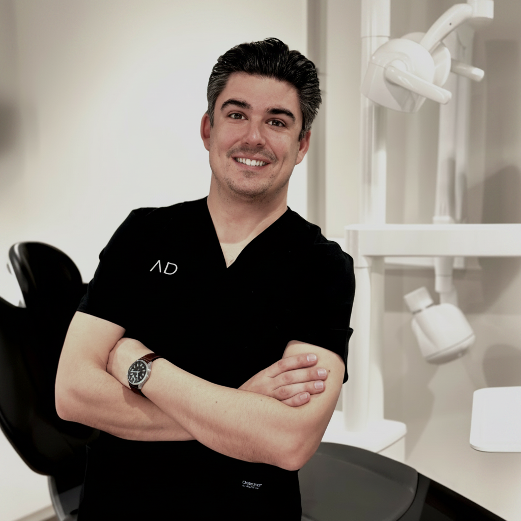
La práctica de Carlos Polis Yanes gira en torno a Cirugía Oral e Implantología. Ejerce en Barcelona. Cursó sus estudios de Odontología en Universidad Europea en 2011.
Con el tiempo, ha sumado formación específica en prótesis y regeneración. Su título de posgrado más reciente es el Máster en Medicina, Cirugía e Implantología Oral, Odontología y Salud Oral (Universitat de Barcelona, 2015).
Actualmente ejerce como cirujano/implantólogo en Clínica dental Beatriz Rubio, Institut Dra. Iriarte y Healthdent en Barcelona y Huesca. Antes desarrolló su actividad en Clínica Universitaria Odontológica UEM.
Además de la clínica, mantiene actividad de docencia en Universitat de Barcelona y Universidad Europea, en programas como Grado / Licenciado en Odontología y Máster de Formación Permanente en Implantología Oral Avanzada.
Su aportación al colectivo: imparte una formación — entre ellas «Taller de Implantología Monobloque» — en Granada y firma 11 publicaciones científicas en revistas como Applied Sciences y Journal of Clinical and Experimental Dentistry.
Explora su trayectoria
Actualización en Prostodoncia, Disfunción Craneomandibular y Alteraciones del Sueño
Curso · Universitat de Barcelona · Barcelona, España
Modular Regeneración Ósea Guiada y Tejidos Blandos
Curso · Osteógenos · Madrid, España
Máster en Medicina, Cirugía e Implantología Oral, Odontología y Salud Oral
Máster · Universitat de Barcelona · Barcelona, España
Especialista en Medicina Oral, Odontología y Salud Oral
Especialista · Universidad Complutense de Madrid · Madrid, España
Programa de Capacitación Clínica
Curso · Universidad Europea · Madrid, España
Diploma en Cirugía Bucal, Odontología y Salud Oral
Experto · Universidad Complutense de Madrid · Madrid, España
Grado / Licenciado en Odontología
Grado · Universidad Europea · Madrid, España
Healthdent · Huesca, España
Clínica dental Aurum · Barcelona, España
Aurum Dental · Barcelona, España
Clínica dental Beatriz Rubio · Barcelona, España
Institut Dra. Iriarte · Barcelona, España
Clínica Universitaria Odontológica UEM · Madrid, España
Profesor asociado · Grado / Licenciado en Odontología
Universitat de Barcelona · Barcelona, España
Profesor · Máster de Formación Permanente en Implantología Oral Avanzada
Universitat de Barcelona · Barcelona, España
Profesor · Máster de Formación Permanente en Cirugía Bucal e Implantología
Universitat de Barcelona · Barcelona, España
Profesor · Grado / Licenciado en Odontología
Universidad Europea · Madrid, España
24–26 de septiembre de 2026 · Granada · Presencial
Cirugía Oral e Implantología · Implantología Monobloque
Applied Sciences
★ Alto impacto · Q1 · IF 3.618Autores · Remei Prat-Riera, Carlos Polis-Yanes, Sonia Egido‐Moreno, Carlos Arranz-Obispo, Beatríz González-Navarro, José López‐López
Background surgical extraction of impacted mandibular third molars is frequently associated with postoperative pain, swelling and trismus. Kinesio Taping (KT) has been proposed as a non-pharmacological adjunct therapy, despite evidence remaining inconclusive. Methods: a systematic review and meta-analysis were conducted according to PRISMA guidelines and registered in PROSPERO (CRD420251120841). PubMed, Scopus, and Web of Science were searched from January 2010 to October 2025. Randomized controlled trials evaluating KT after mandibular third-molar extraction were included. Risk of bias was assessed using the revised Cochrane Risk-of-Bias Tool (RoB 2.0). Results: Ten randomized controlled trials (442 patients) were included. KT significantly reduced postoperative pain in the early postoperative period (MD −2.06 VAS units, 95% CI −3.15 to −0.96; p = 0.0002; I2 = 97%) and at day 7 (MD −0.78, 95% CI −1.52 to −0.05; p = 0.04; I2 = 99%). Facial edema was reduced in the early postoperative period (SMD −1.46, 95% CI −2.47 to −0.45; p = 0.005; I2 = 95%), though estimates should be interpreted with caution given the methodological heterogeneity. No significant reduction was observed at day 7 (SMD −0.33, 95% CI −0.92 to 0.27; p = 0.28; I2 = 87%). Mouth opening improved in both the early (MD −2.78 mm; I2 = 100%) and late periods (MD −4.63 mm; I2 = 87%). The overall certainty of evidence was rated as very low (GRADE). Conclusion: KT may reduce pain and trismus after third-molar extraction, with a reduction in facial swelling. Due to very high heterogeneity, high risk of bias, and very low certainty of evidence, these findings should be interpreted with caution. Further high-quality placebo-controlled trials are needed.
Journal of Clinical and Experimental Dentistry
Q3 · IF 1.368Autores · Carlos Polis-Yanes, Carla Cadenas-Sebastián, S. Egido-Moreno, José López‐López
Background: Bone and soft tissue remodeling after tooth loss is an inevitable physiological process. By placing immediate dental implants and performing a custom healing abutment (CHA), we can help achieve a stable emergence profile and reduce tissue volume loss. Purpose: The purpose of this work is to present a series of two clinical cases to show the Spider Web Technique for the creation of custom healing abutments (CHAs) chair-side in immediate implants. Material and Methods: We present two cases of lower molars with CHAs performed chair-side in the clinic using titanium abutments and flowable composite with the Spider Web Technique as an alternative to prefabricated titanium or polyetheretherketone (PEEK) ones. A brief discussion was held providing other techniques for carrying out CHAs. Results: The creation of CHAs was simple and fast and the postoperative period was very good. After the osseointegration period, the appearance of the peri-implant tissues, as well as the bone level, were optimal for making the definitive crown. Conclusions: The Spider Web Technique provides an effective way to create a CHA around a provisional titanium abutment immediately intraoperatively chair-side with low cost and predictable results. Comparative protocolized studies with others are necessary to obtain clear conclusions and protocols.
Avances en Odontoestomatología
Q4 · IF 0Autores · A Bertran-Faus, Carlos Polis-Yanes, Beatríz González-Navarro, E. Jané‐Salas, Albert Estrugo Devesa, A Marí-Roig, José López‐López
Introducción: Los queratinocitos presentes en las células epiteliales del cuerpo humano producen, de manera continuada, pequeñas cantidades de histamina que se mantienen en equilibrio en el epitelio oral. Cuando este equilibro se ve alterado, se produce un aumento de histamina en el tejido oral pudiendo provocar lesiones.
Medicina oral, patología oral y cirugía bucal
Q2 · IF 2.165Autores · Claudia Oliver-Puigdomenech, Beatríz González-Navarro, Carlos Polis-Yanes, Albert Estrugo‐Devesa, Enric Jané Salas, José López‐López
BACKGROUND: Metastases in the oral cavity are rare and account for only 1 to 3% of all malignant lesions in this area. The primary location from which most metastases have been described in the oral cavity in adult patients include lungs, breasts, kidneys and colon. MATERIAL AND METHODS: A systematic search of the literature was carried out following the PRISMA statement in PubMed database. Clinical trials and case series published in the last 10 years [2010-2020] were eligible to be selected. The headings and keywords used in the searches were "cancer" AND "oral metastases", "incidence" AND "oral metastases", "oral metastases" AND "jaw bone", "oral metastases" AND "soft tissue". RESULTS: For the study of the incidence of metastases in the oral cavity, 9 reports of clinical trials and 7 retrospective studies of case series have been included in this article. The primary locations from which more metastases have been described in the oral cavity are lungs (30.6% or 183 cases), breasts (22.2% or 133 cases), liver (15.5% or 93 cases), prostate (9 % or 54 cases), thyroid glands (8.1% or 49 cases), kidneys (7.3% or 44 cases), skin (2.3% or 14 cases), soft tissues (2% or 12 cases), colon (2% or 12 cases) and gastrointestinal (0.6% or 4 cases). These metastases have a predilection for hard tissues. The clinical presentation of these lesions varies from painless granulomatous lesions to lytic areas in the jaws. CONCLUSIONS: Although metastases in the oral cavity is an uncommon pathology, early diagnosis is needed so that in the event that it is the first manifestation, it allows the primary tumor to be diagnosed as soon as possible.
Book Publisher International (a part of SCIENCEDOMAIN International)
Autores · Carlos Polis-Yanes, Carla Cadenas-Sebastián, Patricia Gual-Vaqués, Raúl Ayuso‐Montero, Antonio Marí-Roig, José López‐López
Alloplastic dental implants are currently the best way to replace lost teeth. In order to achieve good function and prognosis of dental implants, having bone and soft tissue to support them is necessary. When the amount of bone left is not enough to ensure the outcome of the implant, techniques such as shorts implants, zygomatic implants, or guided bone regeneration have been used. Even though autologous bone is mostly the “gold standard,” other biomaterials such as xenografts have led to the reduction of the morbidity of treatments and to the improvement of the regeneration technique outcomes. We present a clinical case of severe atrophy of the maxilla in which we used different types of biomaterials: heterologous cortical lamina, xenograft and autologous bone, and microscrews.
PubMed
Autores · María Apoita-Sanz, Patricia Blanco-Jauset, Carlos Polis-Yanes, Rosa Maria Penin-Mosquera, Gallego Montserrat-Gomá, Laura Pozuelo-Arquimbau, August Vidal-Bell, Carlos Arranz-Obispo, José López‐López
PURPOSE: To present an update the orofacial manifestations of granulomatosis with polyangiitis (GPA) and present a clinical case with the initial signs in the oral cavity. MATERIALS AND METHODS: A bibliographic search was performed on Pubmed with the keywords 'Wegener's granulomatosis', 'etiology', 'oral manifestations', 'oral cavity', 'gingiva'. The inclusion criteria were papers published in English in the last 10 years that made reference to clinical cases with in which the oral cavity was affected. The quality of the results was assessed with 'The 2013 Care Checklist'. RESULTS: Nineteen clinical cases were analysed. The average quality was 7.68/13 (range 5-10/13). 73.7% of patients were women, the most frequent area for the lesions was the gingiva and the most prevalent lesion was gingival hyperplasia. 68.4% of the patients had this lesion as a first sign, 21.1% as a progression and 10.5% as a recurrence. 68.4% of the lesions resolved once medical treatment was established. CONCLUSION: GPA is a multisystem disorder associated with considerable morbidity and mortality if not treated. Early diagnosis improves the prognosis. The first manifestation of the disease can be seen in the oral cavity. It is important that dentists recognise the oral manifestation in order to improve the prognosis. Key words: granulomatosis, polyangiiitis, Wegener's granulomatosis.
Case Reports in Dentistry
Q4 · IF 0.469Autores · Carlos Polis-Yanes, Carla Cadenas-Sebastián, Claudia Oliver-Puigdomenech, Raúl Ayuso‐Montero, Antonio Marí-Roig, José López‐López
When a tooth is extracted, a bone remodeling of the alveolar process occurs irretrievably. Various techniques have emerged over time to maintain the thickness of the bone crest in fixed prosthetics on teeth and implants. The socket shield and pontic shield techniques are aimed at minimizing buccal bone remodeling, especially in the aesthetic area. We present a case of an aesthetic sector rehabilitated with partial fixed denture using the socket shield and pontic shield techniques.
Dipòsit Digital de la Universitat de Barcelona (Universitat de Barcelona)
Autores · António Granate-Marques, Carlos Polis-Yanes, María Seminario-Amez, Enric Jané Salas, José López López
Objetivo: el objetivo de esta revisión es determinar si el tratamiento con Bifosfonatos u otros medicamentos antirresortivos y antiangiogénico influye en el éxito de la regeneración y / o tratamiento implantológico. Material y Métodos: se realizó una revisión bibliográfica en la base de datos PubMed en los últimos 5 años utilizando las siguientes palabras: "Sinus Floor Augmentation"[Mesh] OR "Dental Implants"[Mesh]) OR "Guided Tissue Regeneration"[Mesh]) AND "Osteonecrosis"[Mesh]. Los artículos seleccionados según los criterios de inclusión y exclusión fueron evaluados utilizando los 22 ítems de la declaración STROBE. Aplicamos la siguiente pregunta clínica PICO: ¿influye la terapia con agentes asociados a la Osteonecrosis medicamentosa en el éxito de los tratamientos regeneradores y implantológicos?. Resultados: en la búsqueda inicial se obtuvieron un total de 27 artículos. Tras eliminar los que no hacían referencia al tema, estaban duplicados o no cumplían los criterios de inclusión/exclusión se realizó una lectura completa de los artículos y evaluamos su calidad metodológica, obtuviendo 6 estudios de elevada calidad metodológica y 2 de moderada. Conclusiones: La literatura que trata este tema es escasa, serían necesarios ensayos clínicos randomizados para establecer protocolos de actuación relativos al tratamiento implantológico, en pacientes en tratamientos antirresortivos. El riesgo de desarrollar una Osteonecrosis asociada a la regeneración/colocación de implantes en pacientes con enfermedades óseas benignas y con tratamiento antirresortivo es escaso, pero existe y no debe subestimarse. Sobre todo, en las áreas posteriores de la mandíbula/maxilar, si la duración del tratamiento con bifosfonatos es de más de 3 años, y si el paciente está bajo tratamiento con corticosteroides.
Medicina oral, patología oral y cirugía bucal
Q2 · IF 2.165Autores · A Granate-Marques, Carlos Polis-Yanes, María Seminario-Amez, Enric Jané Salas, José López-López
BACKGROUND: The aim of this study was to determine if the treatment with bisphosphonates other anti-resorptive and antiangiogenic agents influences the success of regenerative and / or implant treatments. MATERIAL AND METHODS: We reviewed the literature from the last 5 years in the PubMed database, using the following words: "Sinus Floor Augmentation"[Mesh] OR "Dental Implants"[Mesh]) OR "Guided Tissue Regeneration"[Mesh]) AND "Osteonecrosis"[Mesh]. The articles were selected following the inclusion and exclusion criteria and were evaluated using the 22 items of the STROBE declaration. The following PICO clinical question was applied: Does the treatment with agents associated with drug osteonecrosis influence the success of regenerative and implant treatments? RESULTS: The initial search resulted in a total of 27 articles. After eliminating those that did not refer to the topic, were duplicated or did not meet the inclusion / exclusion criteria, a full reading of the articles was made evaluating their methodological quality, obtaining six studies with high methodological quality and two with moderate. CONCLUSIONS: The literature regarding this topic is scarce, randomized clinical trials would be necessary to establish protocols relative to implant treatment in patients on antiresorptive treatments. The risk of developing an osteonecrosis associated with the regeneration / implant placement in patients with benign bone diseases is scarce, but it exists and it should not be underestimated. Especially, in the posterior areas of the jaw, if the duration of treatment with BP is greater than 3 years, and if the patient is under therapy with systemic corticosteroids.
Case Reports in Dentistry
Q4 · IF 0.469Autores · Carlos Polis-Yanes, Carla Cadenas-Sebastián, Patricia Gual-Vaqués, Raúl Ayuso‐Montero, Antonio Marí-Roig, José López‐López
Alloplastic dental implants are currently the best way to replace lost teeth. In order to achieve good function and prognosis of dental implants, having bone and soft tissue to support them is necessary. When the amount of bone left is not enough to ensure the outcome of the implant, techniques such as shorts implants, zygomatic implants, or guided bone regeneration have been used. Even though autologous bone is mostly the "gold standard," other biomaterials such as xenografts have led to the reduction of the morbidity of treatments and to the improvement of the regeneration technique outcomes. We present a clinical case of severe atrophy of the maxilla in which we used different types of biomaterials: heterologous cortical lamina, xenograft and autologous bone, and microscrews.
Medicina oral, patología oral y cirugía bucal
Q2 · IF 2.165Autores · Patricia Gual-Vaqués, Carlos Polis-Yanes, Albert Estrugo‐Devesa, Raúl Ayuso‐Montero, Antonio Marí-Roig, José López‐López
BACKGROUND: Recently, bone graft materials using permanent teeth have come to light, and clinical and histological outcomes of this material have been confirmed by some studies. The aim of this systematic review was to evaluate the reliability of the autogenous tooth bone graft material applied to alveolar ridge augmentation procedures. MATERIAL AND METHODS: A systematic review of literature was conducted analyzing articles published between 2007 and 2017. The following four outcome variables were defined: a) implant stability b) post-operative complication c) evaluation of implant survival and failure rates, and d) histological analysis. A total of 108 articles were identified; 6 were selected for review. Based on the PICO (problem, intervention, comparison, outcome) model, the chief question of this study was: Can patients with alveolar ridge deficiency be successfully treated with the autogenous teeth used as bone graft? RESULTS: The mean primary stability of the placed implants was 67.3 ISQ and the mean secondary stability was 75.5 ISQ. The dehiscence of the wound was the most frequent complication with a rate of 29.1%. Of the 182 analyzed implants, the survival rate was 97.7% and the failure rate was 2.3%. In the histological analysis, most of studies reported bone formation. CONCLUSIONS: There is insufficient evidence regarding the effects of autogenous teeth used for bone grafting to support any definitive conclusions, although it has been shown clinically safe and good bone forming capacity, and good results are shown about implant stability.
Cirugía Oral e Implantología
Madrid · España
Cirugía Oral e Implantología
Madrid · España
Cirugía Oral e Implantología
Madrid · España
Cirugía Oral e Implantología
Chile
Cirugía Oral e Implantología
Málaga · España
Cirugía Oral e Implantología
Madrid · España