Cirugía Oral e Implantología
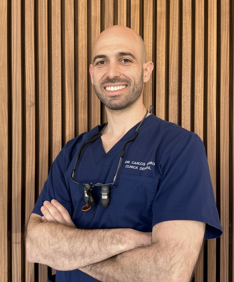
Carlos M. Cobo Vázquez centra su práctica en Cirugía Oral e Implantología. Ejerce en Madrid. Cursó sus estudios de Odontología en Universidad Complutense de Madrid en 2011.
Con el tiempo, ha sumado formación específica en prótesis y ha llegado al nivel de doctorado. Su título de posgrado más reciente es el Doctor en Odontología (Universidad Complutense de Madrid, 2024).
Actualmente ejerce como odontólogo general en Hospital General Universitario Gregorio Marañón y Cobo & Garrido Odontología Especializada en la provincia de Madrid.
Además de la clínica, mantiene actividad de docencia en Universidad Complutense de Madrid, en programas como Grado / Licenciado en Odontología y Postgrado de Especialización en Cirugía Bucal e Implantología.
Su aportación al colectivo: ha publicado 3 casos clínicos sobre implantología, cirugía oral y estética dental y firma 23 publicaciones científicas en revistas como Acta Odontologica Scandinavica e International Journal of Implant Dentistry.
Explora su trayectoria
Doctor en Odontología
Doctorado · Universidad Complutense de Madrid · Madrid, España
Doctor en Ciencias Biomédicas
Doctorado · Universidad Complutense de Madrid · Madrid, España
Especialista en Implantoprótesis
Especialista · Universidad Complutense de Madrid · Madrid, España
Máster en Cirugía Oral e Implantología
Máster · Universidad Complutense de Madrid · Madrid, España
Licenciado en Odontología
Grado · Universidad Complutense de Madrid · Madrid, España
Hospital General Universitario Gregorio Marañón · Madrid, España
Cobo & Garrido Odontología Especializada · Madrid, España
Profesor asociado · Grado / Licenciado en Odontología
Universidad Complutense de Madrid · Madrid, España
Profesor colaborador · Postgrado de Especialización en Cirugía Bucal e Implantología
Universidad Complutense de Madrid · Madrid, España
 9 imgs
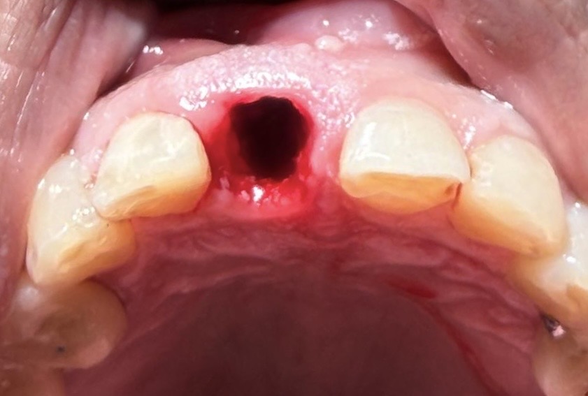
Fractura radicular vertical de un incisivo superior resuelta con exodoncia atraumática, colocación inmediata del implante en posición palatina y relleno del gap vestibular con biomaterial.
 6 imgs
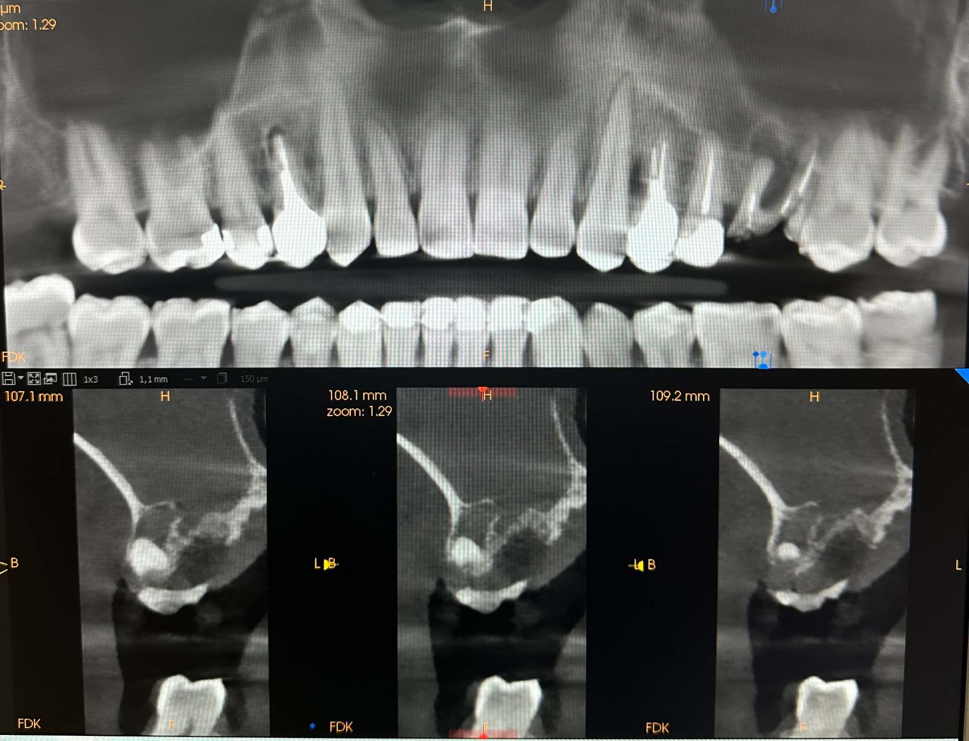
Exodoncia de un resto radicular del 26 con patología periapical y sinusitis asociada, y cierre de la comunicación bucosinusal con bola adiposa de Bichat para preservar el fondo de vestíbulo.
 4 imgs
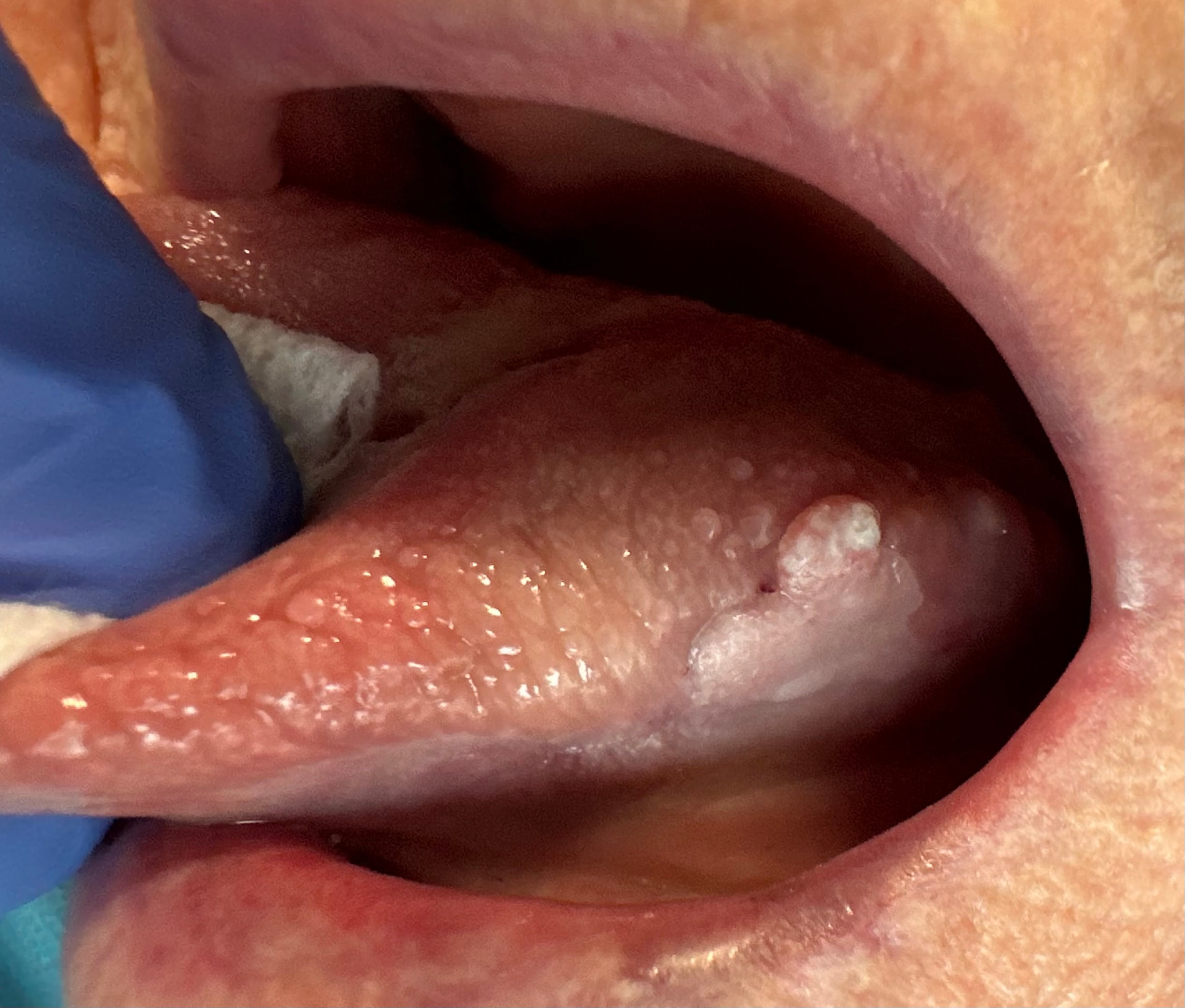
Lesión blanca en borde lateral izquierdo de la lengua con sospecha de leucoplasia en paciente exfumadora polimedicada. La biopsia incisional confirma un carcinoma escamoso microinfiltrante.
Acta Odontologica Scandinavica
Q2 · IF 1.733Autores · Lucía Hernando-Calzado, Aida Bauer-González, Carlos Cobo-Vázquez, Cristina Méniz-García, Juan López-Quiles, Cristina Madrigal Martínez-Pereda
BACKGROUND: Medication-related osteonecrosis of the jaw (MRONJ) is an adverse condition in patients receiving antiresorptive or antiangiogenic therapies. Standard treatments, including surgical debridement, often yield suboptimal outcomes. In this context, bone morphogenetic proteins (BMPs), have been explored for their ability to stimulate osteogenesis and enhance bone repair. Materials and methods: A systematic review of the literature was conducted, focusing on studies that applied rh-BMPs during surgeries to treat MRONJ. Databases were searched for relevant articles from inception to the present, using keywords such as 'MRONJ', 'BMP', and 'bone regeneration'. Inclusion criteria involved studies with human participants who had been treated with rh-BMPs, along with the surgical elimination of bone sequestrum, MRONJ stages 2 and 3 according to the AAOMS staging system and a minimum follow-up period of 6 months. Two independent reviewers (L.H.C. and C.C.V.) systematically selected the articles independently. RESULTS: The review included nine studies with a total of 217 patients treated with rh-BMP. Bone regeneration and osteonecrosis healing was reported in all the studies included using rh-BMP. However, the measurement methods were very different between the studies, using clinical examinations, different radiological tests and biomarkers and own scales. Moreover, there were inconsistencies in treatment protocols and follow-up periods, making it difficult to standardize conclusions. DISCUSSION: While rh-BMPs show promising results for bone regeneration in MRONJ patients, the variability in study methodologies limits definitive conclusions. The biological potential of BMPs could be beneficial, but standardized protocols and longer-term studies are needed to establish their effectiveness. CONCLUSIONS: The application of rh-BMPs may promote bone regeneration in MRONJ patients, but further research with standardized methods is required to confirm these findings.
International Journal of Implant Dentistry
★ Alto impacto · Q1 · IF 4.405Autores · Carlos Cobo-Vázquez, Sonia García-Rodríguez, María Eugenia Colmenares-Otero, Luis Miguel Sáez-Alcaide, Jorge Cortés‐Bretón Brinkmann, Cristina Madrigal Martínez-Pereda, Cristina Méniz-García
PURPOSE: Maxillary sinus floor elevation is a safe and effective surgical technique for achieving vertical bone height, performed through either a lateral or crestal approach. The latter includes both the osteotome technique and osseodensification. The aim of this systematic review was to compare the outcomes of the classic crestal sinus lift technique and the osseodensification sinus lift approach in terms of the bone gain, marginal bone loss, survival rate, follow-up time and complications. METHODS: This review was performed following PRISMA guidelines. An electronic search was conducted across three databases: (1) The National Library of Medicine (MEDLINE/PubMed); (2) SCOPUS; and (3) Cochrane Central Register of Controlled Trials (CENTRAL). The Newcastle-Ottawa Quality Assessment Scale and the Cochrane Collaboration tool for evaluating risk of bias. A meta-analysis for random effects was carried out for implant survival, residual bone height and bone gain. RESULTS: Thirteen studies were included, ten studies performed the osteotome (OST) approach and three performed the osseodensification (OD) approach, with a total of 519 sites treated. The residual bone height was 5.94 and 5.00 mm for OD and OST, respectively. For bone gain, similar results were found for both groups, being 3.37 mm for OD and 3.18 mm for OST. For both groups, the most used diameter and length of the implant was 4 and 10 mm, respectively, and the implant survival rates ranged from 94.1% to 100%. OST technique reflected a complication rate of 14.32%, compared to the OD technique, which showed a complication rate of 2.78%. CONCLUSIONS: It can be concluded that the maxillary sinus lift by osseodesinfication approach is a safe, predictable and successful technique compared to the osteotome approach, with similar outcomes regarding bone gain which is an important parameter for implant placement.
Research Square
Autores · Carlos Cobo-Vázquez, Pedro Molinero‐Mourelle, Cristina Madrigal-Martínez-Pereda, Luis Sánchez‐Labrador, Cristina Méniz-García, José María Martínez González, Juan López-Quiles
Objectives : The main objective was to determine the effectiveness of melatonin in the treatment of peri-implantitis. Secondary objectives were to determine the anti-inflammatory, osteogenic and antimicrobial properties of melatonin. Materials and Methods : A randomized clinical trial was carried out in patients affected by peri-implantitis with a follow-up of 60 days. Melatonin 1.9 mg or placebo was applied locally to the peri-implant defect after debridement. The probing depth, bleeding index, plaque index, and interleukin-1β and 6 concentrations, peri-implant bone level and the bacterial strains were analyzed. Results : 30 patients were analyzed with a mean age of 71.9±7.3 years. Changes on the probing depth in the melatonin group were -1.80±0.88 mm and -1.38±0.47 mm in placebo. Bone level changes were -0.87±0.74 mm in the melatonin group and -0.80±0.68 mm in the placebo. The interleukin-1β concentration at 60 days was 8.62±5.09 pg/mL in the melatonin group and 9.07±5.57 pg/mL in placebo. No differences were observed in probing depth (p=.17), plaque index (p=.57), bleeding index (p=.91), peri-implant bone level (p=.43), interleukin-1β (p=.80), interleukin-6 (p=.52) and anaerobic bacterial species (p=0.96). There were differences in the P. gingivalis concentration (p=.05). Conclusions : Anti-inflammatory, analgesic, osteogenic and antimicrobial properties of melatonin cannot be determined. It seems to improve slightly probing depth changes, interleukin-1β levels and P. gingivalis concentration. Trial registration : Protocol Registration & Results System Clinical Trial Number NCT06816277- Clinical Relevance: Melatonin could improve plaque index, bleeding index, reduce probing depth and peri-implant marginal bone loss, in addition to controlling certain bacterial species in the short term.
Journal of Oral and Maxillofacial Research
Q3 · IF 1.317Autores · Aida Bauer-González, José Bartolomé-Lechuga, Javier Sanz-Alonso, Carlos Cobo-Vázquez, Juan López-Quiles, Cristina Madrigal Martínez-Pereda
Objectives: The primary objective of this systematic review is to evaluate the effectiveness of photobiomodulation in promoting neurosensory regeneration in patients with inferior alveolar nerve injury resulting from the extraction of mandibular third molars or dental implant placement. Material and Methods: An electronic search was conducted across major scientific databases including MEDLINE (PubMed), Scopus, Web of Science, and the Cochrane Library. Predefined inclusion and exclusion criteria were applied, and a total of 10 studies were included for qualitative analysis. Results: A total of 10 studies were analysed, including 4 randomized clinical trials, 5 case series, and 1 retrospective study. All utilized GaAlAs lasers for photobiomodulation (PBM) therapy with wavelengths ranging from 808 to 830 nm. The number of treatment sessions ranged from 7 to 20, delivered at frequencies of 1, 2, or 3 times per week. Nearly all studies reported statistically significant improvements in both objective and subjective neurosensory assessments in favour of PBM therapy. Additionally, no significant differences were found between groups regarding sex, age, injury aetiology, or time to treatment initiation. Conclusions: Photobiomodulation shows promising potential in the neurosensory recovery of inferior alveolar nerve injuries. However, further randomized controlled trials with larger sample sizes and long-term follow-up are needed to establish standardized clinical protocols and confirm its efficacy in dental practice.
Dentistry Journal
★ Alto impacto · Q1 · IF 3.289Autores · Luis Sánchez‐Labrador, Santiago Bazal-Bonelli, Fabián Pérez‐González, Tomás Beca-Campoy, Carlos Cobo-Vázquez, Jorge Cortés‐Bretón Brinkmann, José María Martínez González
Background/Objectives: Subperiosteal implants (SPIs) were first used in the 1940s, but due to their complications and the rise of dental implants, they were discontinued. Thanks to new technologies and new materials, nowadays they are being used again and studied as a treatment for severe bone defects. This review analyzes the clinical results—survival rates and complications—of SPIs used to support full arch rehabilitations of severely resorbed maxillae and mandibles, comparing the outcomes resulting from implant placement conducted in one or two surgical interventions. Methods: An automated search was conducted in four databases (Medline/Pubmed, Scopus, Web of Science, and Cochrane Library), as well as a manual search for relevant clinical articles published before 28 February 2025. The review included human studies with at least four patients, in which SPIs were placed to restore full-arch edentulous maxillae and mandibles. Quality of evidence was evaluated using the Newcastle–Ottawa Quality Assessment Scale and the Joanna Briggs Institute Critical Appraisal tool. Results: A total of 14 studies met the inclusion criteria and were included for analysis, including 958 patients and 973 SPIs. The survival rate was 100% when one surgical intervention was performed and 85% when two interventions were performed after 4–38 months and 3–22 years follow-up, respectively. Conclusions: SPIs would appear to offer a good alternative for patients with severe bone atrophies, especially SPIs fabricated using digital techniques in a single step, presenting promising survival rates and a low complication rate, although more randomized clinical trials with long-term follow-up are needed.
Biomimetics
★ Alto impacto · Q1 · IF 4.339Autores · Diba Ghodsian, Sofía D’Jesús, Luis Sánchez‐Labrador, Carlos Cobo-Vázquez, Jorge Cortés‐Bretón Brinkmann, José María Martínez González, Cristina Méniz-García
The aim of this systematic review was to determine whether autogenous tooth grafting material (ATGM) is as safe and effective as other bone substitutes used for maxillary sinus augmentation procedures, evaluating histomorphometric and/or histological data, implant primary stability, associated complications and radiographic bone height measurements. An automated electronic search was conducted using four databases (Medline/PubMed, Scopus, Web of Science and Cochrane Library), supplemented by a manual search, to identify clinical human studies using particulate ATGM for the aforementioned procedure. The included studies had a sample size of at least four patients and were published before 31st July 2024. The Newcastle-Ottawa scale (NOS) and Joanna Briggs Institute (JBI) Critical Appraisal Checklist were used to assess the risk of bias in cohort studies and case series, respectively. Seven studies were included in the descriptive analysis, obtaining 128 participants (46.8% only treated with ATGM) and 192 placed implants. Due to the heterogeneity of the studies, meta-analysis could not be performed. The authors concluded that ATGM appears to be a feasible and safe alternative for maxillary sinus augmentation procedures. These results should be interpreted with caution due to the limited amount of scientific evidence on this topic and the heterogeneity between the included studies.
Journal of Dentistry
★ Alto impacto · Q1 · IF 4.94Autores · Álvaro Limones, Gülce Çakmak, Manrique Fonseca, Andrea Roccuzzo, Carlos Cobo-Vázquez, Miguel Gómez‐Polo, Pedro Molinero‐Mourelle
PURPOSE: To assess the impact of involuntary interruptions (simulating tracking loss by moving the scanner out of its focal distance) and voluntary interruptions (pressing the scanner's turn-on button) on the accuracy of implant-supported full-arch scans using an intraoral scanner (TRIOS 5, version 22.1.10; 3Shape; Copenhagen, Denmark). MATERIALS AND METHODS: An edentulous model with four implants was digitized with an industrial scanner (Artec Micro II; Artec 3D) to create a reference scan. Four groups (n = 30) were established based on the number of interruptions during scanning: Zero Group (no interruptions; control group), 6-V Group (six voluntary interruptions), 6-I Group (six involuntary interruptions), and 12-I Group (twelve involuntary interruptions). Primary outcome was accuracy assessed by the Root Mean Square (RMS) method. Secondary outcomes included scanning time and the number of photograms. Data were analyzed using one-way ANOVA and post hoc Tukey multiple comparison tests (α=0.05). RESULTS: A total of 120 digital scans were conducted. The Zero group achieved a RMS error of 291 ± 47 µm, a scanning time of 68 ± 6s, and 1320 ± 129 photograms. 6-V group significantly reduced RMS error (MD -102 µm [IC 95 %: -141, -63]), decreased scanning time (MD -20s [IC 95 %: -25, -17]), and reduced photograms (MD -415 photograms [IC 95 %: -506, -324]) compared to the control group (P<.001). Simulations of 6 or 12 involuntary interruptions did not affect accuracy compared to the control group (P>.05). CONCLUSIONS: Voluntary interruptions during scanning, achieved by pressing the scanner's turn-on button, appear to enhance accuracy due to image preprocessing, while involuntary interruptions had no significant impact on the accuracy of implant-supported full-arch scans. CLINICAL SIGNIFICANCE: Voluntary stop during scanning implant-supported full-arches may result in better-fitting prostheses owing to higher scan accuracy and efficiency.
Journal of Dental Anesthesia and Pain Medicine
Q2 · IF 1.908Autores · Carlos Cobo-Vázquez, Milena Gasco
Background: Excessive fear of dental procedures leads to disruptive behavior during dental examinations and treatments. Dental examinations and treatments of these patients usually require additional techniques, such as sedation. The most commonly used techniques are inhalation of nitrous oxide, infusion of propofol with fentanyl, and premedication and infusion of midazolam. Methods: A prospective observational epidemiological study was conducted on patients who required sedoanalgesia techniques for dental exploration and procedures. The reasons for the inability of patients to cooperate (excessive fear or intellectual disability), age, sex, weight, systemic pathology, oral pathology, treatment performed, time of intervention, anesthetic technique performed, and occurrence of complications were recorded. Results: In total, 218 patients were studied. Sixty-five patients came for fear of dental treatment and 153 for presenting with a diagnosis of intellectual disability and not collaborating in the treatment with local anesthesia. The average age of all patients was 30.54 ± 17.30 years. The most frequent oral pathologies found in patients with excessive fear were tartar (6.8%) and wisdom teeth (6.4%), followed by missing teeth (5%). In patients with disabilities, a combination of tartar and cavities appeared most frequently (41.3%), followed by cavities (15.6%). The most frequently used sedoanalgesia technique was the infusion of propofol with fentanyl in both groups of patients, followed by nitrous oxide. Conclusion: The combination of propofol and fentanyl was the most frequently used alternative in patients who were unable to collaborate because of intellectual disability or carry out longer or more complex treatments. Inhaled nitrous oxide and midazolam were the sedative techniques of choice for simpler oral treatments, such as tartrectomies, shallow obturations, and shorter interventions, or in younger patients.
Journal of Stomatology
Q3 · IF 0.802Autores · Carlos Cobo-Vázquez, Luis Sánchez‐Labrador, David Vielba Dueñas, Luis Miguel Sáez-Alcaide, María Isabel Leco Berrocal, Cristina Méniz-García
A relationship between various specialties and adverse effects of post-transplant treatments gives rise to a permanent search for ways to reduce these complications.Aim of the study was to investigate the guidelines for the mana gement of solid organ transplant patients after oral surgery treatment, considering medication employed.A literature review of scientific articles was carried out with systematic search.Terms used were "Oral Surgery" AND "Solid Organ Transplanted Patient" OR "Organ Transplanted".Medline, Web of Science, and Scopus databases were explored, with no time restriction and in Spanish and English languages, complemented by a manual search.A total of 162 titles were retrieved.Out of these, six underwent full data extraction and were included in analysis.Data of 784 patients, with age ranging from 18 to 80 years, and a history of liver, heart, kidney, or pancreas transplants were investigated.Patients who underwent surgical dental treatment, such as extractions, biopsies, and scaling and root planning, to which different medications were administered were considered.Antibiotic prophylaxis was the most common medication administered, in addition to other medicines applied in the treatment of complications, such as post-operative bleeding.In conclusions, there is not enough scientific evidence to establish clear and universal recommendations for the management of solid organ transplant patients after oral surgery.
Journal of Prosthetic Dentistry
Q2 · IF 2.791Autores · Carlos Cobo-Vázquez, Pedro Molinero‐Mourelle, Marta Romeo Rubio, Blanca Guisado‐Moya, Jaime Del Rìo‐Highsmith, Juan López-Quiles
Short implants are a therapeutic alternative for edentulous patients with severe bone resorption. Differences in peri-implant bone loss and complications of short implants depending on the type of connection are unclear. The main purpose of this clinical study was to evaluate the survival rate after 2 years of the short implants in the Oxtein system (Proclinic). Secondary objectives were to compare implant survival, peri-implant bone loss, peri-implant mucosal status, and associated complications in internal hexagonal connection versus external hexagonal connection implants. A randomized clinical trial was carried out in 14 patients with a mean age of 62.7 ±8.5 years, with a total of 61 Oxtein L35 and L6 Proclinic implants being placed at the Faculty of Dentistry. A descriptive analysis, simple binary logistic regression model using generalized estimating equations and Kaplan-Meier survival analysis were carried out (α=.05). Implant survival was 85.2% (52/61). Failure of all implants occurred before prosthetic loading; bleeding after probing occurred in 28 implants, being greater among external connection implants (57.6%) P=.025. The presence of plaque appeared in 36 of the implants, without statistically significant differences between external connection (72.8%) and internal connection (60.0%) (P>.05). A total of 28 implants had at least 2 mm of keratinized mucosa, without statistically significant differences between external connection (63.6%) and internal connection (35.0%) P=.200. A total of 8 complications (13.1%) were recorded, including connection fractures, screw fracture, framework fracture, and buccal fenestrations. Short implants are a therapeutic solution to more complex surgical techniques. However, as lower survival has been reported, the characteristics of the implants and operator experience are important factors for their success.
Lasers in Dental Science
Q4 · IF 0.533Autores · Elena de Górgolas Fernández-Chacón, Andrea Fernández-Juárez, Carlos Cobo-Vázquez, Luis Sánchez‐Labrador, Fabián Pérez‐González, Cristina Méniz-García
The availability of the new generation of direct-acting antiviral (DAA) therapies for the treatment of chronic hepatitis C virus (HCV) infection has completely transformed the HCV therapeutic landscape. Current regimens provide pan-genotypic coverage that are of short duration, safer, and are highly effective than prior generations of HCV therapeutics. While data from clinical trials and real-world experience continue to demonstrate similar and high sustained virologic response (SVR) rates, the successful implementation of the HCV cascade-of-care including screening, confirmation of active infection, treatment initiation and successful completion of therapy among more challenging populations, such as the underserved populations who are also disproportionally affected by HCV, remains vital to HCV eradication efforts.
Journal of Stomatology Oral and Maxillofacial Surgery
Q2 · IF 1.759Autores · Carlos Cobo-Vázquez, Laura Fernández-Gutiérrez, Borja Pérez-Fernández, Luis Sánchez‐Labrador, Natalia Martínez Rodríguez, José María Martínez González, Cristina Méniz-García
The dentigerous cyst is defined as a pathological cavity whose origin is produced by developmental alterations. Most usual treatment is the enucleation of the cyst with the extraction of the tooth. The current approach tends to preserve the tooth. The aim of this study was to determine the total and partial success of the conservative treatment of the dentigerous cyst in terms of eruption of the associated permanent tooth. Pub-Med, Scopus and the Cochrane Central Register of Controlled Trials were searched from January 2012 to December 2023, including patients aged 18 years old or less with a dentigerous cyst associated with a repositionable permanent tooth in the arch. 118 articles were found and 24 were included in full text. 40 cases were reported in the mandible (83 %) and 8 in the maxilla (17 %). 29 cases of marsupialization were included (60 %), 17 of enucleation (36 %) and 2 of decompression (4 %). The percentage of teeth that erupted spontaneously, either completely or partially, after marsupialization, enucleation and decompression was 83 %, 59 % and 100 % respectively. No recurrences have been described in any case. This study highlights that the conservative treatment was a predictable procedure with a total success of 83 % by marsupialization and 100 % after decompression.
Journal of Stomatology Oral and Maxillofacial Surgery
Q2 · IF 1.759Autores · Luis Miguel Sáez-Alcaide, B. Gallego, Javier Fernando Moreno, Miguel Moreno Navarro, Carlos Cobo-Vázquez, Jorge Cortés‐Bretón Brinkmann, Cristina Méniz-García
Vertical bone augmentation procedures are increasingly necessary in daily practice. However, it has been reported that vertical ridge augmentation is one of the least predictable techniques in terms of complications. The aim of this systematic review was to evaluate and compare complications in relation to the different procedures used for vertical bone augmentation prior to implant placement. This review was conducted according to PRISMA guidelines. An electronic search was carried out in four databases: The National Library of Medicine (MEDLINE/PubMed); Web of Science; SCOPUS; and Cochrane Central Register of Controlled Trials (CENTRAL). The Newcastle-Ottawa Quality Assessment Scale, the Cochrane Collaboration tool for assessing risk of bias, and The Joanna Briggs Institute Critical Appraisal tool were used to assess the quality of evidence in the studies reviewed. Twenty-five studies with a total of 749 vertically augmented sites were included in the review. Complication rates varied among the different procedures: 51.02% for distraction osteogenesis, 38.01% for bone blocks, and 16.80% for guided bone regeneration. Vertical bone augmentation procedures prior to implant placement are associated with frequent surgical complications and should be approached with caution due to their possible impact on clinical treatment success.
Journal of Stomatology Oral and Maxillofacial Surgery
Q2 · IF 1.759Autores · Luis Felipe Burgos-Larraín, Álvaro Vázquez-Portela, Carlos Cobo-Vázquez, Luis Miguel Sáez-Alcaide, Luis Sánchez‐Labrador, Cristina Méniz-García
PURPOSE: Odontogenic infections can spread through different routes to more remote anatomical areas, such as the brain. Brain abscesses have an incidence of 0.3-1.3 / 100,000 population and only 2-5% are of dental origin. The main objective is to research brain complications derived from odontogenic infections. Secondary objectives were to identify the most common symptoms in brain abscess, to describe the microbiology involved in these infectious processes, report which parts of the brain complex are most commonly affected and report the sequelae of this patients. METHODS: A systematic review following the PRISMA Guide and the Joanna Briggs Institute (JBI) Critical Appraisal Checklist for Case Reports was carried out in PubMed, Scopus and Web of Science. The search terms were: Brain infection, brain abscess, oral health oral origin, odont* infect*. RESULTS: The database search identified a total of 1000 articles. A total of 18 publications were identified after applying inclusion and exclusion criteria. A total of 38 patients were analyzed. Mean age was 49.64±18.80 years. CONCLUSION: The most common symptoms of patients with brain abscess are neurological affectations first and then fever and headache second, without necessarily presenting as a symptomatological triad. Microbiological diagnosis is key to determining the origin of the infection. Anaerobic pathogens such as Streptococcus (F. Milleri), Fusobacterium Nucleatum and Porfiromonas Gingivalis families are common bacterial agents. The frontal lobe is the most frequently affected, followed by the parietal and temporal lobe. The most frequent brain complications are neurological disorders. However, most patients with brain abscesses recover without sequelae.
International Journal of Environmental Research and Public Health
★ Alto impacto · Q1 · IF 3.251Autores · Santiago Bazal-Bonelli, Luis Sánchez‐Labrador, Jorge Cortés‐Bretón Brinkmann, Carlos Cobo-Vázquez, Natalia Martínez Rodríguez, Tomás Beca-Campoy, Juan Santos-Marino, Emilio Rodríguez-Fernández, Mario Alvarado-Lorenzo
This systematic literature review set out to investigate the relationship between serum vitamin D levels and dental implants in terms of survival rates, marginal bone loss, and associated complications. The review was conducted according to PRISMA guidelines, performing an electronic search in four databases (Pubmed, Web of Science, Cochrane, and Scopus), complemented by a manual search up to April 2022. Four articles were selected for analysis. The Newcastle-Ottawa Quality Assessment Scale tool was used to assess the quality of evidence of cohort studies, and the Cochrane bias assessment tool was used to assess the quality of evidence of randomized clinical trials. The study included 1089 patients restored with 1984 dental implants, with follow-up periods ranging from 20-240 months. Cases presenting lower serum vitamin D levels obtained slightly worse results in terms of marginal bone loss. Longer follow-up periods are needed in order to determine whether serum vitamin D levels affect implant survival rates and osseointegration over time.
Acta Odontologica Scandinavica
Q2 · IF 1.733Autores · Ana Federica Stran-Lo Giudice, Azahara María Ortiz, Luis Sánchez‐Labrador, Jorge Cortés‐Bretón Brinkmann, Carlos Cobo-Vázquez, Cristina Méniz-García
Objetive. During the last few years, cyanoacrylate has been used for wound closure in oral and maxillofacial surgery with growing frequency. When comparing cyanoacrylate with sutures, some authors report similar experiences, while others have found differences. Some agree on the similar outcomes obtained between cyanoacrylate and sutures, others have registered better effects with cyanoacrylate, and others with sutures. Therefore, the aim of this systematic review (SR) was to evaluate postoperative parameters – pain, swelling, trismus, healing and complications (bleeding and infection) – after lower third molar (LTM) removal using cyanoacrylate compared with sutures. Materials and methods. Electronic and manual literature searches were conducted independently by two reviewers up to March 2022. Results. Four studies met the pre-established inclusion criteria and were included for descriptive analysis. These were controlled clinical trials comparing the effects of cyanoacrylate with sutures in 116 patients and 232 split-mouth cases. Pain and haemostasis were significantly reduced on the cyanoacrylate group, swelling showed the same results on two of the studies analysed, trismus and healing had no significant differences between both groups. Conclusions. Both techniques were found to be effective in terms of wound closure, proposing cyanoacrylate as an effective resource that should be investigated in future research. Nevertheless, the literature on cyanoacrylate is scarce and lacks comparative studies of its outcomes and effects.
Applied Sciences
★ Alto impacto · Q1 · IF 3.618Autores · Marta Amigo-Basilio, Covadonga Álvarez-González, Carlos Cobo-Vázquez, María Isabel Leco Berrocal, Luis Miguel Sáez-Alcaide, Cristina Méniz-García
Objective: The aim of this study is to know the biological therapy drugs that are related to adverse events, what dental treatments are associated with the appearance of these events, their severity, and how they are resolved. Study design: Analysis of cases described in the literature on patients undergoing treatment with biological therapies who have developed adverse effects associated with these drugs. Results: Of the 62 articles reviewed, 49 describe 68 cases of MRONJ, most of which appeared in the jaw and received surgical and/or conservative treatment. Conclusions: Biological therapies can potentially develop adverse effects in the oral cavity, so strict monitoring by the dentist is necessary.
Applied Sciences
★ Alto impacto · Q1 · IF 3.618Autores · Andrea Roccuzzo, Pedro Molinero‐Mourelle, Martina Ferrillo, Carlos Cobo-Vázquez, Luis Sánchez‐Labrador, Antonio Ammendolia, Mario Migliario, Alessandro de Sire
The regeneration of nerve injuries after oral surgery procedures is a quite often attempted procedure in dental medicine. Despite several proposed technical approaches, there is still a lack of consensus on which should be considered the gold standard procedure, even-though in the last decades, the use of collagen-based devices allowing a tension-free direct neurorrhaphy has been used. A systematic search of multiple electronic databases and hand searching was conducted to assess the level of evidence behind the use of type I collagen devices to treat nerve injuries after oral surgery procedures. After screening, four articles (one case series and three retrospective studies) including overall 65 patients suffering from inferior alveolar (IAN)/lingual nerve (LN) injury after mandibular wisdom tooth extraction, met the inclusion criteria and could be included. The Oxford Centre for evidence-based medicine (OCEBM) scaling system was used to evaluate the quality of the included studies. Positive clinical results in terms of sensorial improvements were recorded at least 3 months after surgery, even-though the overall level of evidence is low. The use of collagen membranes to enhance nerve regeneration in oral surgery results in promising results. Nevertheless, additional clinical comparative trials with larger sample sizes are needed.
Científica dental: Revista científica de formación continuada
Autores · Ana Encinas Ramos, Luis Miguel Sáez-Alcaide, Carlos Cobo-Vázquez, Cristina Meniz García
espanolLa extraccion del tercer molar inferior es el procedimiento mas frecuente dentro del campo de la cirugia bucal, siendo el dano del nervio dentario inferior una de las complicaciones mas frecuentes en la extraccion. Como alternativa para disminuir el riesgo de aparicion de esta complicacion surge la coronectomia, tecnica que consiste en la eliminacion de la corona dentaria manteniendo intactas las raices dentro del hueso alveolar. El objetivo del presente articulo fue realizar una puesta al dia sobre la coronectomia en terceros molares inferiores, analizando indicaciones, contraindicaciones, complicaciones y exito de esta tecnica. La coronectomia parece ser una alternativa eficaz a la extraccion para la prevencion de trastornos neurosensoriales en terceros molares impactados en estrecha relacion con el NDI. Sin embargo, es necesaria la realizacion de mas estudios comparando la extraccion convencional con la coronectomia, con un mayor seguimiento, para conocer el exito y las complicaciones a largo plazo de este tratamiento. EnglishThe lower third molar extraction is one of the most common procedures in the field of oral surgery. Furthermore, the damage of the inferior alveolar nerve is one of the most frequent complications related to this procedure. The coronectomy was introduced in 1984, as an alternative approach that tended to reduce the occurrence of this complication. The main objective of this article is to review current evidence of coronectomy applied to mandibular third molars, analyzing indications, contraindications, complications and success of the technique. The coronectomy seems to be an effective alternative to conventional third molar extraction, for the prevention of neurosensorial disorders in impacted third molars in direct relation with inferior alveolar nerve. However, further studies are needed which compare conventional approach with coronectomy with a greater follow-up to understand the long-term morbidities and success of the technique
The International Journal of Prosthodontics
Q3 · IF 1.419Autores · Pedro Molinero‐Mourelle, Alexandra Helm, Carlos Cobo-Vázquez, Walter Yu Hang Lam, Luís Azevedo, Ehn Pow, Miguel Gómez‐Polo
PURPOSE: To systematically review the current evidence on clinical and patient-reported outcomes of implant-supported palatal obturator prostheses. MATERIALS AND METHODS: An electronic search of the PubMed, Web of Science, and Cochrane databases was carried out in June 2019. The titles and abstracts of all articles were screened by two independent reviewers. The references of the subsequently selected studies were further screened for potential articles. Assessment of the selected full texts was performed independently according to established inclusion and exclusion criteria. The quality of the selected studies was determined using the Newcastle-Ottawa scale. Interrater agreement on study selection was calculated using Cohen kappa statistic. RESULTS: The search yielded a total of 2,797 records. Ten studies were selected for data extraction, with a Cohen kappa value of 0.856. Five studies were prospective, and five were retrospective. The survival rates for conventional implants ranged from 21.42% to 100%, whereas for zygomatic implants, the survival rates varied from 30% to 100%. Four studies reported prosthodontic complications, with screw loosening being the most common. Patient quality of life (QoL) was analyzed in six studies. CONCLUSION: In spite of the limitations of the present review, it can be concluded that the clinical outcomes are acceptable in terms of survival rates, implant and prosthodontic complications, and QoL associated with implant-supported maxillary obturator prostheses. QoL of implant-supported prostheses in these patients are acceptable. The general study design was not homogenous between studies.
Autores · Juan López-Quiles Martínez, Carlos Cobo-Vázquez, Laura Baca González, Alexandra Helm González
En el sistema de adquisicion de competencias actual resulta complejo la transmision de habilidades practicas. La metodologia de ensenanza de Cirugia Bucal debe realizarse mediante una metodologia audiovisual adecuada a los tiempos y recursos actuales.
Anesthesia Progress
Q4 · IF 0.43Autores · Carlos Cobo-Vázquez, Gemma De Blas, Pablo García-Canas, María del Carmen Gasco-García
Cardiopulmonary resuscitation requires the provider to adopt positions that could be dangerous for his or her spine, specifically affecting the muscles and ligaments in the lumbar zone and the scapular spinal muscles. Increased fatigue caused by muscular activity during the resuscitation could produce a loss of quality and efficacy, resulting in compromising resuscitation. The aim of this study was to evaluate the maximum time a rescuer can perform uninterrupted chest compressions correctly without muscle fatigue. This pilot study was performed at Universidad Complutense de Madrid (Spain) with the population recruited following CONSORT 2010 guidelines. From the 25 volunteers, a total of 14 students were excluded because of kyphoscoliosis (4), lumbar muscle pain (1), anti-inflammatory treatment (3), or not reaching 80% of effective chest compressions during the test (6). Muscle activity at the high spinal and lumbar (L5) muscles was assessed using electromyography while students performed continuous chest compressions on a ResusciAnne manikin. The data from force exerted were analyzed according to side and muscle groups using Student's t test for paired samples. The influence of time, muscle group, and side was analyzed by multivariate analyses ( p ≤ .05). At 2 minutes, high spinal muscle activity (right: 50.82 ± 9.95; left: 57.27 ± 20.85 μV/ms) reached the highest values. Activity decreased at 5 and 15 minutes. At 2 minutes, L5 activity (right: 45.82 ± 9.09; left: 48.91 ± 10.02 μV/ms) reached the highest values. After 5 minutes and at 15 minutes, activity decreased. Fatigue occurred bilaterally and time was the most important factor. Fatigue began at 2 minutes. Rescuers exert muscular countervailing forces in order to maintain effective compressions. This imbalance of forces could determine the onset of poor posture, musculoskeletal pain, and long-term injuries in the rescuer.
Medicina oral, patología oral y cirugía bucal
Q2 · IF 2.165Autores · Carlos Cobo-Vázquez, Isabel F. Tresguerres, R. Ortega-Aranegui, Juan López-Quiles
OBJECTIVES: The purpose of this study was to assess the anti-inflammatory, analgesic and osteogenic early effects of melatonin on post-extraction sockets of patients requiring third molars extraction. STUDY DESIGN: A randomized, triple-blind clinical trial was made using a split-mouth design. Both lower third molars of 10 patients were extracted and 3 mg of local melatonin or placebo were applied. Concentrations of interleukin-6 and nitrotyrosine were determined on samples of the clot from the socket by independent ELISA tests. Radiographic bone density was evaluated by measuring Hounsfield Units in panoramic and cross sections obtained by digital scanner. Statistical analysis by Kolmogorov-Smirnov test was performed for ELISA data. Bone density was analyzed by Shapiro-Wilk test. Subsequently t test was applied. P<0.05 was considered to be significant. RESULTS: The concentration of interleukin-6 increased with the application of melatonin without statistically significance (361.32 ± 235.22 pg/ml vs 262.58 ± 233.92 pg/ml). Nitrotyrosine concentrations showed values below to the detectability pattern (<0.001 nM) in Optic Density curve. Bone density in panoramic sections at socket after melatonin application showed no significant difference (561.98 ± 105.92 HU vs 598.82 ± 209.03 HU). In cross sections, bone density in the alveolar region showed no significant difference(377.42 ± 125.67 HU vs 347.56 ± 97.02 HU). CONCLUSIONS: Within the limitations of this pilot study, no differences with the application of melatonin were found in terms of the concentration of interleukin-6 and bone density in post-extraction socket of retained mandibular third molars.
Cirugía Oral e Implantología
Madrid · España
Cirugía Oral e Implantología
Madrid · España
Cirugía Oral e Implantología
Zaragoza · España
Cirugía Oral e Implantología
Madrid · España
Cirugía Oral e Implantología
Madrid · España
Cirugía Oral e Implantología
Zaragoza · España