Cirugía Oral e Implantología
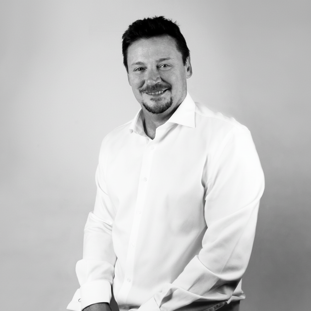
Osmundo Gilbel del Águila centra su práctica en Cirugía Oral e Implantología, y trabaja de forma específica la cirugía avanzada y regenerativa. Ejerce en Murcia. Se formó como odontólogo en Universidad de Murcia en 2015.
Su recorrido formativo explica el resto: ha orientado su formación de posgrado hacia periodoncia y ha llegado al nivel de doctorado. Su título de posgrado más reciente es el Doctorado en Odontología (Universidad de Murcia, 2015).
Actualmente ejerce como cirujano/implantólogo en Clínica dental Europa, Clínica dental Raúl Navarro y Fisiodent Azarbe en la provincia de Murcia. Antes desarrolló su actividad en Clínica dental Dr. Laureano Hernández y Clínica dental Enéresi.
Compagina la clínica con la docencia en Universidad de Murcia, en programas como Máster en Implantología y biomateriales.
Dentro de Doctor&Cols firma 5 publicaciones científicas en revistas como Journal of Functional Biomaterials y BMC Oral Health.
Explora su trayectoria
Doctorado en Odontología
Doctorado · Universidad de Murcia · Murcia, España
Especialista universitario en Periodoncia
Especialista · Universidad de Murcia · Murcia, España
Grado en Odontología
Grado · Universidad de Murcia · Murcia, España
Clínica dental Europa · Murcia, España
Clínica dental Raúl Navarro · Murcia, España
Fisiodent Azarbe · Murcia, España
Unión dental Sierra Minera · Murcia, España
Clínica dental Implantologika · Murcia, España
Clínica dental Arquesi Odontología · Murcia, España
Hospital USP San Carlos · Murcia, España
Clínica dental Dr. Laureano Hernández · Albacete, España
Clínica dental Enéresi · Murcia, España
Clínica dental Ferrando · Murcia, España
Profesor · Máster en Implantología y biomateriales
Universidad de Murcia · Murcia, España
Journal of Functional Biomaterials
★ Alto impacto · Q1 · IF 5.284Autores · Fabio Camacho-Alonso, Osmundo Gilbel-Del Águila, Paula Ferrer-Díaz, David Peñarrocha-Oltra, Yolanda Guerrero-Sánchez, Juan Carlos Bernabeu-Mira
This study involved a randomized clinical trial that included 140 patients. Alveolar ridge preservation was performed with xenografts. Sealing in the control group consisted of a collagen membrane versus cyanoacrylate in the test group. The dental implants were placed immediately after extraction. The variables were evaluated at 3, 12, and 18 months of follow-up. Pearson's chi-squared test was used for qualitative variables and the Student t-test for related samples was used for quantitative variables. The change in buccolingual alveolar bone width was significantly greater in the CMX group than in the CX group after three months (p < 0.005). However, significance was not reached at the other follow-up timepoints (p > 0.005). CAL showed significantly greater values in the CMX group than in the CX group (p < 0.005), and MBL proved greater in the CMX group than in the CX group, with p < 0.001. Five membrane exposures were recorded in the CMX group. Cyanoacrylate as a sealing method for alveolar ridge preservation seems to afford better clinical and radiological results than collagen membrane.
BMC Oral Health
★ Alto impacto · Q1 · IF 3.535Autores · Sofía Soler-Alcaraz, Yolanda Guerrero Sánchez, Osmundo Gilbel del Águila, Juan Carlos Bernabeu-Mira, et al.
Sinus lift surgeries are part of the daily practice of dentists. This study evaluates the long-term structure of the bone placed in sinus lifts through the fractal dimension. We conducted a retrospective study on a sample of 35 patients with 51 sinus lifts performed using a lateral window approach and filling material placement. We radiologically analyzed the graft bone to observe its evolution up to one and a half years after the surgical procedure. The obtained results were the average area of the sinuses analyzed was 1401.96 mm2, with a mean area occupied by the filling material of 297.75 mm2. Significant differences are observed when comparing the fractal dimension values obtained on the initial day and one year after prosthesis loading. Similarly, when comparing the values of the area occupied by the biomaterial at the start day and one year after prosthesis loading, significant differences are also obtained (p-value < 0.001). In conclusion, the filling material used in the lateral window sinus lift procedure undergoes significant resorption and shows changes in the fractal dimension.
Medicina Oral, Patología Oral y Cirugía Bucal
Q2 · IF 2.165Autores · D Muñoz-Cámara, O Gilbel-Del Águila, G Pardo-Zamora, F Camacho-Alonso
Background: Few studies have reported the outcomes of immediate placement at infected post-extraction sites. The aim of this study was to compare clinical and radiological outcomes of immediately placed implants with immediate prosthetic provisionalization in sockets with or without acute periapical pathology. Material and methods: A total of 100 patients with immediately placed implants with immediate provisionalization and 1-year of follow up were included (50 patients with acute periapical pathology and a control group of 50 patients without acute periapical pathology). Clinical parameters (bleeding on probing, buccal keratinized mucosa width, clinical recession, and probing depth) and radiological parameters (distance from implant shoulder to first point of bone-to-implant contact [IS-BIC]) were assessed. Results: Clinical parameters showed no significant differences between the study and control groups after 1-year follow up (p>0.05). IS-BIC presented the following values: 0.35 ± 0.51 mm (study group) and 0.15 ± 0.87 mm (control), without significant differences between the groups (p=0.160). None of the 50 radiographs of immediate implants placed in sockets with periapical pathology revealed retrograde peri-implantitis. Conclusions: Immediate placement of implants with immediate prosthetic provisionalization at sites with acute periapical pathology can be a successful treatment modality for at least 1-year.
The International Journal of Periodontics & Restorative Dentistry
Q3 · IF 1.136Autores · Osmundo Gilbel del Águila; Ana Arques Fernández; Álvaro Ferrando Cascales
Coautores Doctor&Cols · Ana Arques Fernández
The success of oral restorations supported by dental implants in regenerated bone is well documented, as is the use of bone grafts to augment deficient alveolar bone. This article describes the bone-retention wedge graft, which may be useful for increasing the primary stability of dental implants, preserving bone in postextraction sockets, and covering exposed implant threads.
Clinical Implant Dentistry and Related Research
★ Alto impacto · Q1 · IF 3.62Autores · Fabio Camacho-Alonso, Mariano Sánchez-Siles, Osmundo Gilbel-del Águila
Background: Titanium is the most widely used metal in implant dentistry. In spite of its biocompatibility, when it is released into the oral environment, it can have local negative biological effects. Purpose: The aims of this study were to detect the concentration of metal ions in patients with dental implants, to evaluate whether or not their release might be influenced by the presence of other metals, and to assay whether these ions might provoke genotoxic damage in oral mucosa cells. Materials and methods: One hundred five patients with a total of 180 dental implants were included. The sample was divided into seven groups (n = 15 per group). Group 1 consisted of patients with metal-porcelain fixed crowns on dental implants; Group 2, patients with metal-porcelain fixed crowns on teeth; Group 3, patients with dental amalgams; Group 4, patients with metal-porcelain fixed crowns on dental implants and metal-porcelain fixed crowns on teeth; Group 5, patients with metal-porcelain fixed crowns on dental implants and dental amalgams; and Group 6, patients with metal-porcelain fixed crowns on dental implants, metal-porcelain fixed crowns on teeth, and dental amalgams. Group 7 was the control group, without any dental treatment. The concentration of metal ions was detected using inductively coupled plasma mass spectrometry; genotoxicity was measured using the buccal micronucleus cytome assay protocol. Results: Group 5 displayed the highest concentration of metal ions in parts per billion (Ti, Co, Ni, Zn, Pd, Sn, and Pb). Group 6 was characterized by the highest presence of Hg. No signs of genotoxic damage were found in any of the study groups. Conclusions: Patients with titanium dental implants combined with other metal restorations presented higher concentrations of metal ions, but no genotoxic damage was observed in oral mucosal epithelial cells. Keywords: dental implants; genotoxic damage; metal ions.
Cirugía Oral e Implantología
Málaga · España
Cirugía Oral e Implantología
Madrid · España
Cirugía Oral e Implantología
Madrid · España
Cirugía Oral e Implantología
Madrid · España
Cirugía Oral e Implantología
Las Palmas · España
Cirugía Oral e Implantología
Valencia · España